

Hemorrhage and large lipid-rich necrotic cores are independently associated with thin or ruptured fibrous caps: an in vivo 3T MRI study
- PMID: 19608971
- PMCID: PMC2750883
- DOI: 10.1161/ATVBAHA.109.192179
Hemorrhage and large lipid-rich necrotic cores are independently associated with thin or ruptured fibrous caps: an in vivo 3T MRI study
Abstract
Objective: Histological studies suggest associations between hemorrhage and large lipid-rich/necrotic cores with thin or ruptured fibrous caps in advanced atherosclerosis. We investigated these associations in carotid arteries with mild to severe stenosis by in vivo 3T MRI.
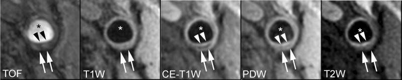
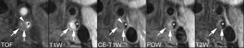
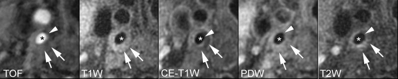
Methods and results: Seventy-seven patients with >or=50% carotid stenosis in at least one side by duplex ultrasound underwent bilateral multi-contrast carotid MRI scans. Measurements for wall and lipid-rich/necrotic core sizes, presence of hemorrhage, and fibrous cap status (classified as intact thick, intact thin or ruptured) were recorded. Arteries with poor image quality, occlusion, or no detectable lipid-rich/necrotic core were excluded. For the 798 MRI slices included, multivariate ordinal regression analysis demonstrated larger %lipid-rich/necrotic core (odds ratio for 10% increase, 1.49; P=0.02) and presence of hemorrhage (odds ratio, 5.91; P<0.001) were independently associated with a worse (intact thin or ruptured) stage of fibrous cap status. For artery-based multivariate analysis, a larger maximum %lipid-rich/necrotic core and presence of hemorrhage independently associated with worse fibrous cap status (P<0.001, for both). No hemorrhage was detected in arteries with thick fibrous caps.
Conclusions: Hemorrhage and larger %lipid-rich/necrotic core were independently associated with a thin or ruptured fibrous cap status at an early to advanced stage of carotid atherosclerosis.
Figures
References
-
- Virmani R, Burke AP, Kolodgie FD, Farb A. Pathology of the thin-cap fibroatheroma: a type of vulnerable plaque. Journal of Interventional Cardiology. 2003;16:267–72. - PubMed
-
- Spagnoli LG, Mauriello A, Sangiorgi G, Fratoni S, Bonanno E, Schwartz RS, Piepgras DG, Pistolese R, Ippoliti A, Holmes DR., Jr Extracranial thrombotically active carotid plaque as a risk factor for ischemic stroke. JAMA. 2004;292:1845–52. - PubMed
-
- Virmani R, Burke AP, Kolodgie FD, Farb A. Vulnerable plaque: the pathology of unstable coronary lesions. Journal of Interventional Cardiology. 2002;15:439–46. - PubMed
-
- Burke AP, Virmani R, Galis Z, Haudenschild CC, Muller JE. 34th Bethesda Conference: Task force #2--What is the pathologic basis for new atherosclerosis imaging techniques? J Am Coll Cardiol. 2003;41:1874–86. - PubMed
-
- Virmani R, Kolodgie FD, Burke AP, Farb A, Schwartz SM. Lessons from sudden coronary death: a comprehensive morphological classification scheme for atherosclerotic lesions. Arterioscler Thromb Vasc Biol. 2000;20:1262–75. - PubMed
Publication types
MeSH terms
Grants and funding
LinkOut - more resources
Full Text Sources
Other Literature Sources
Medical
Miscellaneous
