

National Institutes of Health Stroke Scale score is poorly predictive of proximal occlusion in acute cerebral ischemia
- PMID: 19608992
- PMCID: PMC2763301
- DOI: 10.1161/STROKEAHA.109.555664
National Institutes of Health Stroke Scale score is poorly predictive of proximal occlusion in acute cerebral ischemia
Abstract
Background and purpose: Multimodal imaging is gaining an important role in acute stroke. The benefit of obtaining additional clinically relevant information must be weighed against the detriment of increased cost, delaying time to treatment, and adverse events such as contrast-induced nephropathy. Use of National Institutes of Health Stroke Scale (NIHSS) score to predict a proximal arterial occlusion (PO) is suggested by several case series as a viable method of selecting cases appropriate for multimodal imaging.
Methods: Six hundred ninety-nine patients enrolled in a prospective cohort study involving CT angiographic imaging in acute stroke were dichotomized according to the presence of a PO, including a subgroup of 177 subjects with middle cerebral artery M1 occlusion.
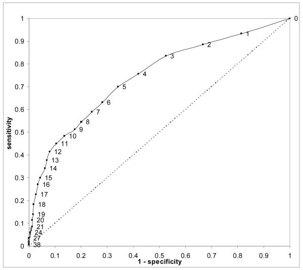
Results: The median NIHSS score of patients found to have a PO was higher than the overall median (9 versus 5, P<0.0001). The median NIHSS score of patients with middle cerebral artery M1 occlusion was 14. NIHSS score > or =10 had 81% positive predictive value for PO but only 48% sensitivity with the majority of subjects with PO presenting with lower NIHSS scores. All patients with NIHSS score > or =2 would need to undergo angiographic imaging to detect 90% of PO.
Conclusions: High NIHSS score correlates with the presence of a proximal arterial occlusion in patients presenting with acute cerebral ischemia. No NIHSS score threshold can be applied to select a subgroup of patients for angiographic imaging without failing to capture the majority of cases with clinically important occlusive lesions. The finding of minimal clinical deficits should not deter urgent angiographic imaging in otherwise appropriate patients suspected of acute stroke.
Figures


References
-
- Saver JL. Time is brain--quantified. Stroke. 2006;37(1):263–266. - PubMed
-
- Grotta J. NIHSS/EIC mismatch explains the >1/3 MCA conundrum. Stroke. 2003;34(9):e148–9. author reply e148-9. - PubMed
-
- Barber PA, Darby DG, Desmond PM, Yang Q, Gerraty RP, Jolley D, Donnan GA, Tress BM, Davis SM. Prediction of stroke outcome with echoplanar perfusion- and diffusion-weighted MRI. Neurology. 1998;51(2):418–426. - PubMed
-
- Butcher K, Parsons M, Baird T, Barber A, Donnan G, Desmond P, Tress B, Davis S. Perfusion thresholds in acute stroke thrombolysis. Stroke. 2003;34(9):2159–2164. - PubMed
-
- Parsons MW, Li T, Barber PA, Yang Q, Darby DG, Desmond PM, Gerraty RP, Tress BM, Davis SM. Combined (1)H MR spectroscopy and diffusion-weighted MRI improves the prediction of stroke outcome. Neurology. 2000;55(4):498–505. - PubMed
Publication types
MeSH terms
Grants and funding
LinkOut - more resources
Full Text Sources
Other Literature Sources
Medical
