. 2009 Sep;66(17):2933-42.
doi: 10.1007/s00018-009-0082-7. Epub 2009 Jul 16.

Differential use of an in-frame translation initiation codon regulates human mu opioid receptor (OPRM1)

Affiliations

Differential use of an in-frame translation initiation codon regulates human mu opioid receptor (OPRM1)

Kyu Young Song et al. Cell Mol Life Sci. 2009 Sep.

Abstract

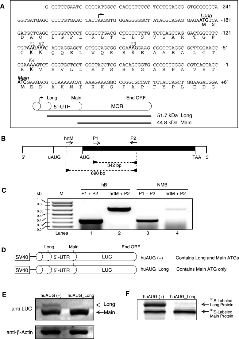
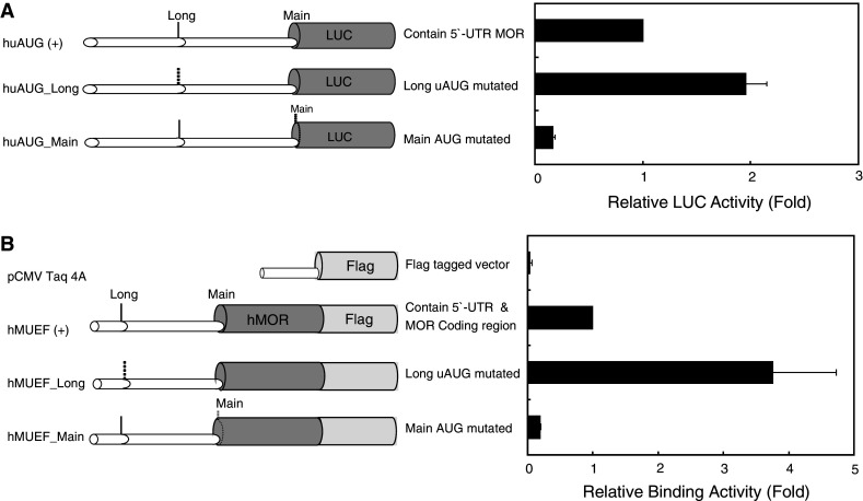
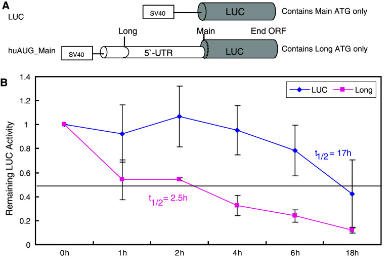
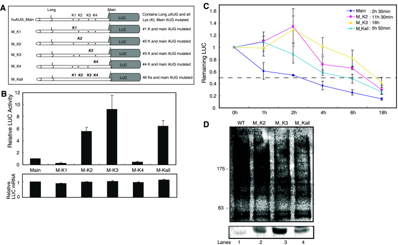
The pharmacological effects of morphine and morphine-like drugs are mediated primarily through the micro opioid receptor. Here we show that differential use of an in-frame translational start codon in the 5'-untranslated region of the OPRM1 generates different translational products in vivo and in vitro. The 5'-end of the OPRM1 gene is necessary for initiating the alternate form and for subsequent degradation of the protein. Initiation of OPRM1 at the upstream site decreases the initiation at the main AUG site. However, alternative initiation of the long form of OPRM1 produces a protein with a short half-life, resulting from degradation mediated by the ubiquitin-proteasome pathway. Reporter and degradation assays showed that mutations of this long form at the second and third lysines reduce ubiquitin-dependent proteasome degradation, stabilizing the protein. The data suggest that MOP expression is controlled in part by initiation of the long form of MOP at the alternate site.


Figures

Fig. 1
Fig. 1
Schematic representation of the human μ opioid receptor (OPRM1) and putative 5′-UTR sequences. a Sequence of the OPRM1 5′-UTR used in this study. The in-frame uAUG (Long) and main AUG (Main) codons are indicated, as are the peptide sequences for the lysine (K1, K2, K3, and K4) codons. The arrowhead above the sequence indicates the transcription initiation site. The upstream and main AUGs encode the Long and Main forms of OPRM1, respectively. b Reverse transcription (RT)-PCR was carried out using oligonucleotide primers (“Materials and methods”). The diagram shows the relative position of sense (hrtM and P1; forward arrows) and antisense (P2; reverse arrows) primers along the OPRM1 mRNA. The darkened area represents the exon region (1,403 bp). Vertical lines represent the relative positions of the common 5′ end, initiation codon for the Long form of OPRM1 (uAUG), initiation codon for the Main form of OPRM1 (AUG), termination codon for both OPRM1s (TAA), and the 3′ end. c RT-PCR analysis of the OPRM1. Total RNA prepared from human brain (hB, lanes 1 and 2; Ambion) and NMB cells were subjected to RT-PCR with the oligonucleotide primer pairs indicated above the lanes followed by agarose gel electrophoresis. Sizes of amplification products obtained with the indicated primer pairs are shown. M denotes DNA markers, the sizes of which are shown on the left. d Luciferase fusion constructs containing portions of OPRM1 extension sequences. The huAUG (+) construct retains both the 5′ in-frame and main AUGs; the huAUG_Long construct retains only the main AUG. The dotted line represents ATGs converted to ACGs by point mutations. e NMB cells were transfected transiently with the constructs shown in d, and protein levels were analyzed by Western blotting for luciferase and β-actin. f Representative autoradiogram of proteins translated in vitro in the presence of [35S]-methionine using a coupled transcription/translation system
Fig. 2
Fig. 2
Translation of the OPRM1 gene is controlled by upstream AUG initiation. a Schematic representations of reporter constructs with wild-type and mutant human 5′-UTRs. Dotted lines represent ATGs converted to ACGs by point mutations. Relative LUC activity in NMB cells transfected transiently with the constructs (right panel). Bars indicate the standard errors of triplicate LUC assays. b Schematic representations of constructs containing the OPRM1 promoter and coding regions. The coding regions were fused in-frame to the FLAG tag, except for the negative control vector (pCMV-Taq4A). [3H]diprenorphine-binding assays performed on opioid receptor-expressing NMB cells transfected transiently with the constructs (right panel). Data represent the mean binding ± standard errors from representative triplicate assays
Fig. 3
Fig. 3
The 5′-extended long form of MOP is a short-lived protein. a Schematics of the constructs used. The initiation codon (dotted line) indicates a point mutation of ATG to ACG. b Time course of protein degradation. The relative remaining LUC activity in NMB cells transfected transiently with the constructs shown in a was determined by incubating the cells in serum-free medium at 37°C for 0–18 h. Cell lysates were assayed for luciferase reporter activity, expressed as the ratio LUC/β-gal. Data represent the means and standard errors of three independent assays conducted in duplicate. t 1/2 = half-life
Fig. 4
Fig. 4
Effects of protease inhibitors on the degradation of the long form of MOP. NMB cells were transfected for 24 h with the constructs shown in Fig. 2b. Cells were then incubated with or without inhibitor at 37°C for 0–12 h, as indicated. The time course of receptor binding was determined as described in Fig. 2. Mean values ± standard errors from representative triplicate assays are shown. a Receptor binding in cells expressing the long form of MOP (hMUEF_Main) incubated with DMSO alone (i.e., no protease inhibitors). b Receptor binding in cells expressing the long form of MOP incubated with 25 μM MG132, leupeptin, or E64. c Receptor binding in cells expressing both the long and main forms of MOP [hMUEF (+)], incubated with 25 μM MG132
Fig. 5
Fig. 5
Mutation of OPRM1 lysines significantly affects the protein’s stability in NMB cells. a Schematic representations of OPRM1 constructs with 5′ lysines (K). Mutated lysines are shown in bold and italicized. b Relative LUC activity and mRNA levels (expressed as the ratios LUC/β-gal and LUC/LacZ, respectively) in NMB cells transfected transiently with each mutant construct. Bars indicate the standard errors of triplicate LUC assays. c Time course of protein degradation. The relative remaining LUC activity in NMB cells transfected transiently with the constructs shown in a was determined as described in Fig. 3b. d NMB cells cultured in six-well plates were transfected with 2 μg of wild-type (lane 1) or mutated (lanes 2–4) constructs. Forty-eight hours after transfection, immunoprecipitation was performed with anti-luciferase and analyzed by immunoblot using either the antibody to ubiquitin (upper panel) or the antibody to luciferase (lower panel). Numbers on the left (kDa) indicate position of molecular weight marker

Similar articles

Cited by

References

    1. Chaturvedi K, Christoffers KH, Singh K, Howells RD. Structure and regulation of opioid receptors. Biopolymers. 2001;55:334–346. doi: 10.1002/1097-0282(2000)55:4<334::AID-BIP1006>3.0.CO;2-S. - DOI - PubMed
    1. Min BH, Augustin LB, Felsheim RF, Fuchs JA, Loh HH. Genomic structure analysis of promoter sequence of a mouse μ opioid receptor gene. Proc Natl Acad Sci USA. 1994;91:9081–9085. doi: 10.1073/pnas.91.19.9081. - DOI - PMC - PubMed
    1. Kieffer B. Recent advances in molecular recognition and signal transduction of active peptides: receptors for opioid peptides. Cell Mol Neurobiol. 1995;15:615–635. doi: 10.1007/BF02071128. - DOI - PMC - PubMed
    1. Kieffer B. Opioids: first lessons from knockout mice. Trends Pharmacol Sci. 1999;20:19–26. doi: 10.1016/S0165-6147(98)01279-6. - DOI - PubMed
    1. Kim CS, Hwang CK, Choi HS, Song KY, Law PY, Wei LN, Loh HH. Neuron-restrictive silencer factor (NRSF) functions as a repressor in neuronal cells to regulate the mu opioid receptor gene. J Biol Chem. 2004;279:46464–46473. doi: 10.1074/jbc.M403633200. - DOI - PubMed

Publication types

MeSH terms