. 2010 Jan;136(1):99-113.
doi: 10.1007/s00432-009-0641-1.

Preclinical characterization of Aurora kinase inhibitor R763/AS703569 identified through an image-based phenotypic screen

Affiliations

Preclinical characterization of Aurora kinase inhibitor R763/AS703569 identified through an image-based phenotypic screen

John McLaughlin et al. J Cancer Res Clin Oncol. 2010 Jan.

Abstract

Purpose: Aurora kinases play a key role in mitotic progression. Over-expression of Aurora kinases is found in several human cancers and correlated with histological malignancy and clinical outcomes. Therefore, Aurora kinase inhibitors should be useful in the treatment of cancers.

Methods: Cell-based screening methods have an advantage over biochemical approaches because hits can be optimized to inhibit targets in the proper intracellular context. We developed a novel Aurora kinase inhibitor R763/AS703569 using an image-based phenotypic screen. The anti-proliferative effect was examined in a panel of tumor cell lines and primary cells. The efficacy was determined in a broad panel of xenograft models.

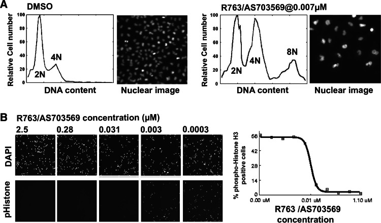
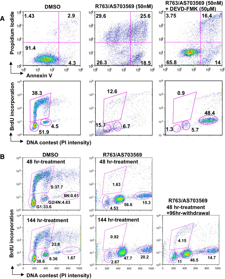
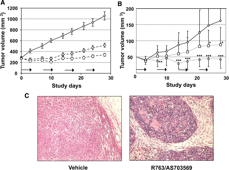
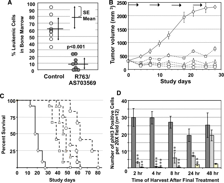
Results: R763/AS703569 inhibits Aurora kinases, along with a limited number of other kinases including FMS-related tyrosine kinase 3 (FLT3), and has potent anti-proliferative activity against many cell types accompanying unique phenotypic changes such as enlarged cell size, endoreduplication and apoptosis. The endoreduplication cycle induced by R763/AS703569 was irreversible even after the compound was withdrawn from the culture. Oral administration of R763/AS703569 demonstrated marked inhibition of tumor growth in xenograft models of pancreatic, breast, colon, ovarian, and lung tumors and leukemia. An acute myeloid leukemia cell line MV4-11, which carries a FLT3 internal tandem duplication mutation, is particularly sensitive to R763/AS703569 in vivo.

Conclusions: R763/AS703569 is a potent inhibitor of Aurora kinases and exhibited significant anti-proliferative activity against a wide range of tumor cells both in vitro and in vivo. Inhibition of Aurora kinases has the potential to be a new addition to the treatment of cancers.


Figures

Fig. 1
Fig. 1
a Induction of enlarged nuclei and 4N/8N arrest by R763/AS703569. A549 cells were incubated with 0.007 μM R763/AS703569 for 48 h. Nuclear morphology and DNA intensity were captured with an inverted fluorescent microscope after cells were fixed and stained with DAPI. DNA content of each nucleus was plotted and smoothed using the Lowess method for cell cycle analysis. b Inhibition of histone H3 serine 10 phosphorylation by R763/AS703569. A549 cells were synchronized with nocodazole and treated with R763/AS703569 for 1 h (0.0001–2.5 μM, 10 points, threefold dilution). The cells were fixed and stained with FITC-labeled anti-phospho-histone H3 serine 10 antibody and DAPI. The number of cells, cell cycle profile, and phospho-histone H3 serine 10 positive cells were measured in the image-based assay. The images obtained at 0.0003, 0.003, 0.031, 0.28, and 2.5 μM are shown in the figure
Fig. 2
Fig. 2
Inhibition of tumor colony formation in vitro by R763/AS703569. Cells from a wide variety of solid tumor xenografts that were directly derived from human primary tumors and maintained in nude mice at Oncotest Inc. (open circles), and xenografts of ATCC-derived tumor cells (closed circles), were used for the clonogenic assays. Each circle represents an EC50 value of each cell line. The mean EC50 value, 10× mean value, and 0.1× mean value are indicated as horizontal bars. ATCC-derived cell lines used in this assay are SF268 glioblastoma (EC50 = 1.185 μM), HT29 colon adenocarcinoma (EC50 = 0.027 μM), K562 chronic myelogenous leukemia (EC50 = 0.010 μM), A549 lung carcinoma (EC50 = 0.004 μM), L363 plasma cell leukemia/lymphoma (EC50 = 0.007 μM), RAJI Burkitt’s lymphoma (EC50 = 0.017 μM), MCF7 breast carcinoma (EC50 = 3.7 μM), MDA-MB-231 breast carcinoma (EC50 = 0.024 μM), MX1 breast carcinoma (EC50 = 0.005 μM), DU-145 prostate carcinoma (EC50 = 0.024 μM), and MRIH1579 prostate carcinoma (EC50 = 0.005 μM)
Fig. 3
Fig. 3
a Inhibition of R763/AS703569-mediated cell death by a caspase-3 inhibitor, DEVD-FMK. Colo205 cells were cultured in 50 nM R763/AS703569 containing media for 48 h with or without 50 μM DEVD-FMK. An apoptosis assay (annexin V/PI staining) and BrdU cell cycle analysis (anti BrdU antibody/PI staining) were performed to assess the effect of R763/AS703569. Percentage of cells in each gate is shown at the corner of the quadrant or next to the gate. 0.05% dimethyl sulfoxide (DMSO) (vehicle) was used for a negative control. b Irreversible cell cycle arrest at 4N and 8N DNA content in A549 cells. Cells were treated with R763/AS703569 for 48–144 h or for 48 h followed by incubation with R763/AS703569 free media for 24–96 h. Percentage of cells within each gate is indicated next to the gate. 0.05% DMSO (vehicle) was used for a negative control
Fig. 4
Fig. 4
Potent anti-tumor activity of R763/AS703569 against human solid tumor cell lines in vivo. Mice were orally treated consecutively for 3 days with various doses of R763/AS703569, followed by a 4-day rest interval. This cycle was repeated four times (shown as arrows). In all animal studies, a 4-week treatment with R763/AS703569 was well tolerated with a minimum body weight loss at the highest dose (less than 10% in all experiments). At the end of the four cycles (including the last 4 day resting), gross and microscopic examination of different tissues from the R763/AS703569-treated mice showed lack of any significant alterations. Toxicity profiling of all tested tissues, including bone marrow and gastrointestinal tract, confirmed that there was no difference between orally treated mice and mice treated through the intra-peritoneal route. No sign of mechanism-independent toxicity of the compound was observed. a SCID mice bearing MiaPaCa-2 pancreatic cancer cells were treated with vehicle control (rhombus, solid line), R763/AS703569 15 mg/kg day (circle, dashed line) or R763/AS703569 20 mg/kg day (pentagon, dashed line). Data are shown as mean tumor volume ± standard error of measurement (SEM) (small bar) of 10 mice/group. b SCID mice bearing NCI-MDR tumors were treated with vehicle (rhombus, solid line), R763/AS703569 7 mg/kg day (open squares, dashed line), or R763/AS703569 10 mg/kg day (open triangles, dashed line). Data are shown as mean tumor volume ± SEM (small bar) of 10 mice/group. Two out of ten mice (the 7 mg/kg day dosing group), and seven out of ten mice (the 10 mg/kg day dosing group) showed clear reduction of tumor volume less than the original volume. The two-sample t test was applied to evaluate the data against the control. Asterisks (*, **, and ***) next to the symbols represent P values of <0.05, <0.01, and <0.001, respectively. c Histological analysis of R763/AS703569-treated tumors. MiaPaCa-2 tumors from mice treated with vehicle or R763/AS703569 20 mg/kg day for 4 weeks were harvested and stained with hematoxylin and eosin
Fig. 5
Fig. 5
Potent anti-tumor activity of R763/AS703569 against human leukemia cell lines in vivo. We used an intermittent schedule of administration as described in materials and methods. a MOLT-4 leukemia cells were implanted i.v. in NOD/SCID mice. Treatment with R763/AS703569 began 14 days after i.v. inoculation of the leukemia cells. The majority of MOLT-4 cells repopulated in bone marrow. Bone marrow cells from NOD/SCID mice bearing Molt4 leukemia cells were harvested after four cycles of the R763/AS703569 treatment. Leukemic cells were identified as human CD45 positive and human CD71 positive cells with a FACScalibur. Data are presented as % leukemic cells in individual mice and mean ± SD (n = 9). The two-sample t test was applied for statistical significance. b MV4-11 leukemia cells, which carry a FLT3 ITD mutation, were injected subcutaneously into NOD/SCID mice. The mice bearing MV4-11 leukemia cells were treated with vehicle control (rhombus, solid line), R763/AS703569 7.5 mg/kg day (open square, dashed line), R763/AS703569 10 mg/kg day (open triangle, dashed line), R763/AS703569 12.5 mg/kg day (circle, dashed line) or R763/AS703569 20 mg/kg day (pentagon, dashed line). The intermittent treatment of R763/AS703569 was repeated four times (shown as arrows). Data are presented as mean tumor volume ± SEM (small bar) (n = 10/group). c Survival of NOD/SCID mice bearing MV4-11 leukemia cells after four rounds of the R763/AS703569 treatment. Mice were killed if body weight loss was >20% of its body weight at the start of drug treatment, if the tumor size of an animal was ≥2,000 mm3, if the tumor ulcerated, or if the mice became moribund. d Reduction of phosphorylated histone H3 expression in MV4-11 xenografts by R763/AS703569. Phospho-histone H3 positive cells were counted and data are presented as the number of phospho-histone H3 positive cells per field (20× magnification) (n = 12 fields per tumor, 3 tumors/group). Hatched bars, dotted bars, and open bars represents mean ± SD of vehicle, R763/AS703569 7.5 mg/kg treated and R763/AS703569 15 mg/kg day treated mice, respectively. The number of phospho-histone H3 positive cells per field in the treated groups of each time point was compared against the vehicle control of the same time point. Asterisks (*, **, and ***) on top of the bars represent P values of <0.05, <0.01, and <0.001, respectively

References

    1. Adams RR, Wheatley SP, Gouldsworthy AM, Kandels-Lewis SE, Carmena M, Smythe C et al (2000) INCENP binds the Aurora-related kinase AIRK2 and is required to target it to chromosomes, the central spindle and cleavage furrow. Curr Biol 10(17):1075–1078 - PubMed
    1. Araki K, Nozaki K, Ueba T, Tatsuka M, Hashimoto N (2004) High expression of Aurora-B/Aurora and Ipll-like midbody-associated protein (AIM-1) in astrocytomas. J Neurooncol 67(1–2):53–64 - PubMed
    1. Barabasz A, Foley B, Otto JC, Scott A, Rice J (2006) The use of high-content screening for the discovery and characterization of compounds that modulate mitotic index and cell cycle progression by differing mechanisms of action. Assay Drug Dev Technol 4(2):153–163 - PubMed
    1. Bayliss R, Sardon T, Vernos I, Conti E (2003) Structural basis of Aurora-A activation by TPX2 at the mitotic spindle. Mol Cell 12(4):851–862 - PubMed
    1. Bischoff JR, Anderson L, Zhu Y, Mossie K, Ng L, Souza B et al (1998) A homologue of Drosophila aurora kinase is oncogenic and amplified in human colorectal cancers. EMBO J 17(11):3052–3065 - PMC - PubMed

MeSH terms

LinkOut - more resources