Comparative Study
. 2009 Sep;18(9):1896-905.
doi: 10.1002/pro.200.

Crystal structure of the variable domain of the Streptococcus gordonii surface protein SspB

Affiliations
Comparative Study

Crystal structure of the variable domain of the Streptococcus gordonii surface protein SspB

Nina Forsgren et al. Protein Sci. 2009 Sep.

Abstract

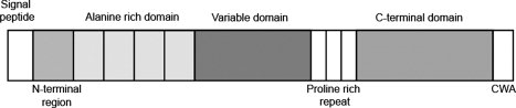
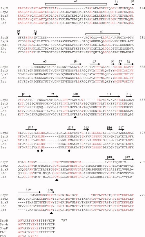
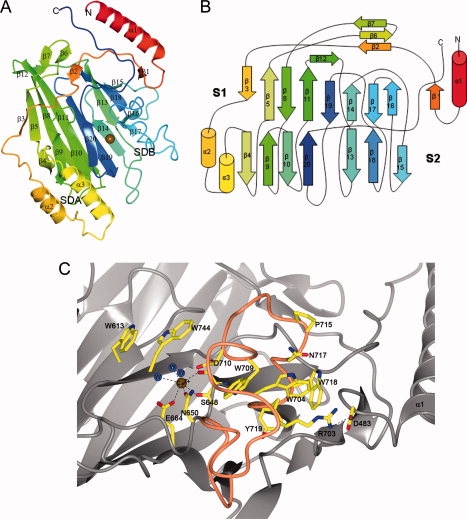
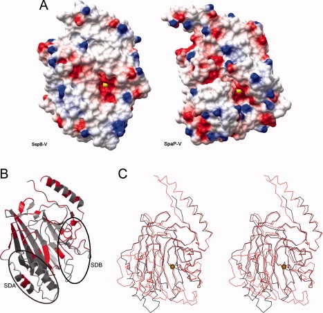
The Antigen I/II (AgI/II) family of proteins are cell wall anchored adhesins expressed on the surface of oral streptococci. The AgI/II proteins interact with molecules on other bacteria, on the surface of host cells, and with salivary proteins. Streptococcus gordonii is a commensal bacterium, and one of the primary colonizers that initiate the formation of the oral biofilm. S. gordonii expresses two AgI/II proteins, SspA and SspB that are closely related. One of the domains of SspB, called the variable (V-) domain, is significantly different from corresponding domains in SspA and all other AgI/II proteins. As a first step to elucidate the differences among these proteins, we have determined the crystal structure of the V-domain from S. gordonii SspB at 2.3 A resolution. The domain comprises a beta-supersandwich with a putative binding cleft stabilized by a metal ion. The overall structure of the SspB V-domain is similar to the previously reported V-domain of the Streptococcus mutans protein SpaP, despite their low sequence similarity. In spite of the conserved architecture of the binding cleft, the cavity is significantly smaller in SspB, which may provide clues about the difference in ligand specificity. We also verified that the metal in the binding cleft is a calcium ion, in concurrence with previous biological data. It was previously suggested that AgI/II V-domains are carbohydrate binding. However, we tested that hypothesis by screening the SspB V-domain for binding to over 400 glycoconjucates and found that the domain does not interact with any of the carbohydrates.


Figures

Figure 1
Figure 1
Domain architecture of Ag I/II proteins. The 1500 residue protein is comprised of seven regions (distinguished by shading): a signal peptide is followed by an N-terminal region, an alanine-rich repeat domain, a variable domain, a proline-rich repeat region, a C-terminal domain and a cell-wall anchoring region (CWA).
Figure 2
Figure 2
Sequence alignment. Multiple sequence alignment of SspB-V and related AgI/II variable domains. Sequences from the following organisms were compared; S. gordonii (SspB); S. gordonii (SspA), S. mutans NG5 (SpaP); S. mutans (PAc) and S. intermedius (Pas). Secondary structure elements of S. gordonii SspB-V are indicated above the sequence. Conserved residues are shown in red. The metal coordinating residues are marked with filled stars and the two conserved tryptophans and a third tryptophan/phenylalanine located in the binding pocket are marked with filled triangles. Numbering is according to the sequence of S. gordonii SspB-V.
Figure 3
Figure 3
Overall structure of SspB-V. (A) Ribbon representation of SspB-V. The coloring blends through the model from red (N-terminus) to blue (C-terminus). The domain is organized into a central immunoglobulin-like β-sandwich flanked by two subdomains, subdomain A (SDA) and subdomain B (SDB). The β-sandwich consists of two anti-parallel β-sheets packed against each other in a β-supersandwich-like fold with a putative binding pocket stabilized by a Ca2+ ion, depicted as a gold sphere. (B) Topology diagram of SspB-V. β-strands are represented as arrows, helices as cylinders and loops as lines. Colors are the same as in (A). (C) The putative binding pocket. The Ca2+ ion (gold sphere) is coordinated by four atoms of the protein and three water molecules (blue spheres). The coiled region comprising the major part of SDB is colored orange. Amino acids in the binding pocket and in SDB are represented as ball-and-stick models and are labeled.
Figure 4
Figure 4
Structure comparison and analysis. (A) Electrostatic surface of SspB-V (left) and SpaP-V, pdb code 1JMM, (right). The molecular surfaces are colored blue and red according to positive and negative electrostatic potential, respectively. The Ca2+ ion is represented by a yellow sphere. (B) Ribbon representation of SspB-V in which the strictly conserved residues are colored red. SDA (Subdomain A) and SDB (Subdomain B) are indicated by oval lines. (C) Stereo view of SspB-V and SpaP-V. SspB-V is colored in black and SpaP-V in red. The Ca2+ ion is represented by a gold sphere. In S. gordonii and S. mutans V-domains, the core of the β-sandwich is highly conserved. The main structural differences reside in SDA resulting in a smaller and shallower pocket in SspB-V.

References

    1. Jenkinson HF, Lamont RJ. Oral microbial communities in sickness and in health. Trends Microbiol. 2005;13:589–595. - PubMed
    1. Nyvad B, Kilian M. Comparison of the initial streptococcal microflora on dental enamel in caries-active and in caries-inactive individuals. Caries Res. 1990;24:267–272. - PubMed
    1. Jenkinson HF, Lamont RJ. Streptococcal adhesion and colonization. Crit Rev Oral Biol Med. 1997;8:175–200. - PubMed
    1. Rosan B, Lamont RJ. Dental plaque formation. Microbes Infect. 2000;2:1599–1607. - PubMed
    1. Douglas CW, Heath J, Hampton KK, Preston FE. Identity of viridans streptococci isolated from cases of infective endocarditis. J Med Microbiol. 1993;39:179–182. - PubMed

Publication types

Substances