. 2010 Jan 15;126(2):426-36.
doi: 10.1002/ijc.24754.

Caveolin-1 promotes resistance to chemotherapy-induced apoptosis in Ewing's sarcoma cells by modulating PKCalpha phosphorylation

Affiliations

Caveolin-1 promotes resistance to chemotherapy-induced apoptosis in Ewing's sarcoma cells by modulating PKCalpha phosphorylation

Oscar M Tirado et al. Int J Cancer. .

Abstract

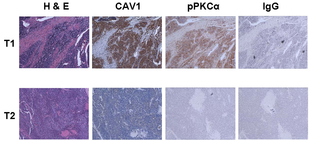
Caveolin-1 (CAV1) has been implicated in the regulation of several signaling pathways and in oncogenesis. Previously, we identified CAV1 as a key determinant of the oncogenic phenotype and tumorigenic activity of cells from tumors of the Ewing's Sarcoma Family (ESFT). However, the possible CAV1 involvement in the chemotherapy resistance commonly presented by an ESFT subset has not been established to date. This report shows that CAV1 expression determines the sensitivity of ESFT cells to clinically relevant chemotherapeutic agents. Analyses of endogenous CAV1 levels in several ESFT cells and ectopic CAV1 expression into ESFT cells expressing low endogenous CAV1 showed that the higher the CAV1 levels, the greater their resistance to drug treatment. Moreover, results from antisense- and shRNA-mediated gene expression knockdown and protein re-expression experiments demonstrated that CAV1 increases the resistance of ESFT cells to doxorubicin (Dox)- and cisplatin (Cp)-induced apoptosis by a mechanism involving the activating phosphorylation of PKCalpha. CAV1 knockdown in ESFT cells led to decreased phospho(Thr(638))-PKCalpha levels and a concomitant sensitization to apoptosis, which were reversed by CAV1 re-expression. These results were recapitulated by PKCalpha knockdown and re-expression in ESFT cells in which CAV1 was previously knocked down, thus demonstrating that phospho(Thr(638))-PKCalpha acts downstream of CAV1 to determine the sensitivity of ESFT cells to chemotherapeutic drugs. These data, along with the finding that CAV1 and phospho(Thr(638))-PKCalpha are co-expressed in approximately 45% of ESFT specimens tested, imply that targeting CAV1 and/or PKCalpha may allow the development of new molecular therapeutic strategies to improve the treatment outcome for patients with ESFT.


Figures

Figure 1
Figure 1
Resistance to Cp and Dox correlates with elevated CAV1 expression levels in ESFT cells. (a) Western immunoblot showing caveolin-1 (CAV1) expression levels in the Ewing’s sarcoma (EWS) and peripheral neuroectodermal tumor (PNET) cell lines indicated; the IC50 values (±SD) of cisplatin (Cp) and doxorubicin (Dox) for each cell line are shown at the bottom. (b) Western immunoblot showing the increased levels of expression attained in TC-71 cells after transfection with a CAV1 cDNA expression construct. In panels a and b, β-actin was used as the loading standard, and the quantitative data shown below the CAV1 lanes were determined by densitometric analysis, normalized relative to the intensity of loading and background signals, and are given as the mean values of three independent experiments. (c) Relative percentage of cell death detected in cultures of untransfected TC-71 cells, cells transfected with the empty cDNA expression vector (TC-71+EV), and cells transfected with the CAV1 cDNA expression construct (TC-71+CAV1) after 24 h treatment with vehicle alone (Control) or with the indicated Cp or Dox concentrations; * P ≤ 0.01 relative to EV-transfected control cells.
Figure 2
Figure 2
CAV1 knockdown sensitizes ESFT cells to Cp- and Dox-induced apoptotic cell death. (a) Western immunoblot showing that transfection with either antisense (AS/CAV1) or shRNA (pShCav1-1) markedly down-regulated the expression levels of caveolin-1 (CAV1) in A4573 cells, relative to cells transfected with the respective control plasmids for the AS (pBK-CMV) or the shRNA (pShCont) constructs; re-expression of CAV1 in cells in which it had been previously knockdown (pShCav1-1+CAV1) using the same cDNA expression construct described for Figure 1 resulted in CAV1 expression levels comparable to those in control cells; quantitative data shown below the CAV1 lanes were determined by densitometric analysis, normalized relative to the intensity of loading and background signals, and are given as the mean values of three independent experiments. (b) Relative percentage of cell death detected in cultures of the same cells described above, after 24 h treatment with vehicle alone (Control) or with the indicated Cp or Dox concentrations; * and ** P ≤ 0.01 relative to their respective controls. (c) Western immunoblot analysis of total extracts prepared from the cells indicated, after treatment (as above) with either vehicle (−) or the indicated drugs (+) to detect the cleavage of XIAP and caspase-3 as apoptosis-specific markers. Wherever applicable, β-actin was used as the loading standard.
Figure 3
Figure 3
Expression of total and Thr638-phosphorylated PKCα in ESFT cells expressing different CAV1 levels. (a) Western immunoblot showing expression levels of total and Thr638-phosphorylated PKCα (pPKCα) in the Ewing’s sarcoma (EWS) and peripheral neuroectodermal tumor (PNET) cell lines indicated; quantitative data shown below the pPKCα and PKCα lanes were determined by densitometric analysis, normalized relative to the intensity of loading and background signals, and are given as the mean values of three experimental replicas. (b) Graphic representation of the densitometric data for CAV1 (from Fig. 1a), and pPKCα and PKCα (from panel a) to compare their relative expression levels in the ESFT cell lines revealed the correlation between the levels of CAV1 and pPKCα, whereas minor variations were detected in total PKCα expression; a value of 1.0 was arbitrarily assigned to the levels of expression of CAV1, total PKCα and pPKCα in A4573 cells, as the cell line with the highest CAV1 content. (c) Western immunoblot showing expression levels of total PKCα and pPKCα in untransfected TC-71 cells and in cells engineered to over-express CAV1 by transfection with the same CAV1 cDNA expression construct described for Figure 1 (TC-71+CAV1); quantitative data shown below the pPKCα and PKCα lanes were determined by densitometric analysis, normalized relative to the intensity of loading and background signals, and are given as the mean values of three experimental replicas. Wherever applicable, β-actin was used as the loading standard.
Figure 4
Figure 4
Inhibition of PKCα sensitizes ESFT cells to Cp- and Dox-induced apoptotic cell death. (a) Relative percentage of cell death detected in cultures of vector transfected A4573 cells (pBK-CMV) and in cultures of A4573 cells in which CAV1 was re-expressed after shRNA-mediated knockdown (pShCav1-1+CAV1) after treatment for 24 h with either the Gö6976 PKCα inhibitor alone, Cp (5 µg/ml) alone, Dox alone (10 ng/ml), or their combinations; * P ≤ 0.01 relative to cells treated with Cp alone. (b) Western immunoblot analysis of total extracts prepared from the cells indicated, after the treatments shown, to detect the expression of cleaved caspase-3 as an apoptosis-specific marker; β-actin was used as the loading standard. (c) Relative percentage of cell death detected in cultures of A4573 cells after treatment for 24 h with either the PKCβ inhibitor (PKCβ inh) alone, Cp (5 µg/ml) alone, Dox alone (10 ng/ml) or their combinations, showing that, contrary to the effect of PKCα inhibition shown in panel a, inhibition of PKCβ did not sensitize the cells to Cp- or Dox-induced apoptosis.
Figure 5
Figure 5
PKCα activation functions downstream of CAV1 to promote drug resistance in ESFT cells. (a) Western immunoblot showing that transfection with either antisense (AS/CAV1) or shRNA (pShCav1-1) caused minor alterations in total PKCα levels, but markedly down-regulated the levels of Thr638-phosphorylated PKCα (pPKCα) in A4573 cells, relative to untransfected cells and to cells transfected with the respective control plasmids for the AS (pBK-CMV) or the shRNA (pShCont) constructs; re-expression of CAV1 in cells in which it had been previously knockdown (pShCav1-1+CAV1), with the same cDNA expression construct described for Figure 1, resulted in the restoration of Thr638-phosphorylated PKCα expression to levels close to those in control cells. (b) Western immunoblot showing that shRNA-mediated knockdown (pShPKCα) markedly down-regulated the expression of PKCα and pPKCα in A4573 cells, relative to untransfected cells and to cells transfected with the control (pShCont) construct; re-expression of PKCα in cells in which it had been previously knocked down (pSh PKCα+PKCα) or in cells in which CAV1 had been previously knocked down efficiently restored the expression of PKCα and pPKCα. (c) Relative percentage of cell death detected in cultures of the cells indicated, after 24 h treatment with vehicle alone (Control) or with Cp, showing that PKCα knockdown sensitized A4573 cells to Cp-induced apoptosis, and that PKCα re-expression in CAV1- and PKCα-knockdown cells restored Cp resistance; * and **, P ≤ 0.01 relative to their respective controls. In panels a and b, β-actin was used as the loading standard, and quantitative data shown below the pPKCα and PKCα lanes were determined by densitometric analysis, normalized relative to the intensity of loading and background signals, and are given as the mean values of three experimental replicas.
Figure 6
Figure 6
Correlation between the expression levels of CAV1 and Thr638-phosphorylated PKCα (pPKCα) in Ewing’s sarcoma tumor specimens. Immunohistochemical analysis for CAV1 and pPKCα in representative tumor samples that either showed positive CAV1 immunostaining (T1) or were negative for CAV1 expression (T2). All but one CAV1 positive tumor samples were also immunoreactive to a pPKCα-specific monoclonal antibody. The specificity of the anti-pPKCα staining was demonstrated by the lack of reactivity with an isotype rabbit IgG control (IgG). H&E, hematoxylin and eosin stained sections. Magnification, ×100.
Figure 7
Figure 7
Model proposed for the mechanism by which the pro-survival function of CAV1 in the response of ESFT cells to drug treatment is mediated by the activating phosphorylation of PKCα at Thr638 rather than by the total cellular levels of PKCα. In ESFT cells with high CAV1 content, CAV1 association with discrete cellular membrane microdomains results in the formation of caveolae, where the high number of CAV1 molecules facilitates the binding of PKCα molecules which are either efficiently activated by phosphorylation at Thr638 upon binding, or remain there more stably in the activated state, if phosphorylated prior to binding. In cells with low CAV1 levels caveolae formation is defective or absent, and CAV1 associates more sparsely with membrane microdomains, thus making the binding, activating phosphorylation and/or the stable maintenance of the phosphorylated status of PKCα less probable, thereby limiting its contribution drug resistance mechanisms. Arrows represent the relative frequency of CAV1–PKCα binding events in the two different cellular contexts.

References

    1. Rodriguez-Galindo C, Spunt SL, Pappo AS. Treatment of Ewing sarcoma family of tumors: current status and outlook for the future. Med Pediatr Oncol. 2003;40:276–287. - PubMed
    1. Barker LM, Pendergrass TW, Sanders JE, Hawkins DS. Survival after recurrence of Ewing's sarcoma family of tumors. J Clin Oncol. 2005;23:4354–4362. - PubMed
    1. Delattre O, Zucman J, Plougastel B, Desmaze C, Melot T, Peter M, Kovar H, Joubert I, de Jong P, Rouleau G, Aurias A, Thomas G. Gene fusion with an ETS DNA-binding domain caused by chromosome translocation in human tumours. Nature. 1992;359:162–165. - PubMed
    1. Bernstein M, Kovar H, Paulussen M, Randall RL, Schuck A, Teot LA, Juergens H. Ewing's sarcoma family of tumors: current management. The Oncologist. 2006;11:503–519. - PubMed
    1. Gupta GP, Massagué J. Cancer metastasis: building a framework. Cell. 2006;127:679–695. - PubMed

Publication types

MeSH terms