Increased expression of ErbB-2 in liver is associated with hepatitis B x antigen and shorter survival in patients with liver cancer
- PMID: 19610068
- PMCID: PMC2745322
- DOI: 10.1002/ijc.24580
Increased expression of ErbB-2 in liver is associated with hepatitis B x antigen and shorter survival in patients with liver cancer
Abstract
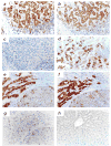
Hepatitis B x antigen, or HBxAg, contributes importantly to the pathogenesis of hepatocellular carcinoma (HCC). Given that HBxAg constitutively activates beta-catenin and that upregulated ErbB-2 promotes beta-catenin signaling in other tumor types, experiments were designed to ask whether HBxAg was associated with upregulated expression of ErbB-2. When HBxAg positive and negative HepG2 cells were subjected to proteomics analysis, ErbB-2 was shown to be upregulated in HepG2X but not control cells. ErbB-2 was also strongly upregulated in HB infected liver, and weakly in some HCC nodules, where it correlated with HBxAg expression. Among tumor bearing patients, strong ErbB-2 staining in the liver was associated with dysplasia, and a shorter survival after tumor diagnosis. This implies that elevated ErbB-2 is an early marker of HCC. Treatment of HepG2X cells with ErbB-2 specific siRNA not only reduced ErbB-2 expression, but also reduced the expression of beta-catenin, suggesting that ErbB-2 contributed to the stabilization of beta-catenin. ErbB-2 specific siRNA also partially blocked the ability of HBxAg to promote DNA synthesis and growth of HepG2 cells. These results suggest that ErbB-2/beta-catenin up-regulation contributes importantly to the mechanism of HBxAg mediated hepatocellular growth.
Conflict of interest statement
There are no conflicts of interest to report.
Figures


Similar articles
-
Enhanced cell survival of Hep3B cells by the hepatitis B x antigen effector, URG11, is associated with upregulation of beta-catenin.Hepatology. 2006 Mar;43(3):415-24. doi: 10.1002/hep.21053. Hepatology. 2006. PMID: 16496348
-
Hepatitis B x antigen up-regulates vascular endothelial growth factor receptor 3 in hepatocarcinogenesis.Hepatology. 2007 Jun;45(6):1390-9. doi: 10.1002/hep.21610. Hepatology. 2007. PMID: 17539024
-
Downregulation of E-cadherin by hepatitis B virus X antigen in hepatocellullar carcinoma.Oncogene. 2006 Feb 16;25(7):1008-17. doi: 10.1038/sj.onc.1209138. Oncogene. 2006. PMID: 16247464
-
Putative roles of hepatitis B x antigen in the pathogenesis of chronic liver disease.Cancer Lett. 2009 Dec 1;286(1):69-79. doi: 10.1016/j.canlet.2008.12.010. Epub 2009 Feb 6. Cancer Lett. 2009. PMID: 19201080 Free PMC article. Review.
-
Hepatitis B virus X antigen in the pathogenesis of chronic infections and the development of hepatocellular carcinoma.Am J Pathol. 1997 Apr;150(4):1141-57. Am J Pathol. 1997. PMID: 9094970 Free PMC article. Review.
Cited by
-
Upregulation of miR-375 inhibits human liver cancer cell growth by modulating cell proliferation and apoptosis via targeting ErbB2.Oncol Lett. 2018 Sep;16(3):3319-3326. doi: 10.3892/ol.2018.9011. Epub 2018 Jun 22. Oncol Lett. 2018. PMID: 30127930 Free PMC article.
-
Common and differential features of liver and pancreatic cancers: molecular mechanism approach.Gastroenterol Hepatol Bed Bench. 2021 Fall;14(Suppl1):S87-S93. Gastroenterol Hepatol Bed Bench. 2021. PMID: 35154607 Free PMC article.
-
Short-chain fatty acids in cancer pathogenesis.Cancer Metastasis Rev. 2023 Sep;42(3):677-698. doi: 10.1007/s10555-023-10117-y. Epub 2023 Jul 11. Cancer Metastasis Rev. 2023. PMID: 37432606 Free PMC article. Review.
-
Prediction of hepatocellular carcinoma risk in patients with chronic liver disease from dynamic modular networks.J Transl Med. 2021 Mar 23;19(1):122. doi: 10.1186/s12967-021-02791-9. J Transl Med. 2021. PMID: 33757544 Free PMC article.
-
Nuclear pore complex protein RANBP2 and related SUMOylation in solid malignancies.Genes Dis. 2024 Sep 4;12(4):101407. doi: 10.1016/j.gendis.2024.101407. eCollection 2025 Jul. Genes Dis. 2024. PMID: 40271196 Free PMC article. Review.
References
-
- Yarden Y, Sliwkowski MX. Untangling the ErbB signaling network. Nat Rev Mol Cell Biol. 2001;2:127–37. - PubMed
-
- Slamon DJ, Clark GM, Wong SG, Levin WJ, Ullrich A, McGuire WL. Human breast cancer: correlation of relapse and survival with amplification of the HER-2/neu oncogene. Science. 1987;235:177–82. - PubMed
-
- Tanner B, Kreutz E, Weikel W, Meinert R, Oesch F, Knapstein PG, Becker R. Prognostic significance of c-erbB-2 mRNA in ovarian cancinoma. Gynecol Oncol. 1996;62:268–77. - PubMed
-
- Weiner DB, Nordberg J, Robinson R, Nowell PC, Gazdar A, Greene MI, Williams WV, Cohen JA, Kern JA. Expression of the new gene-encoded protein (P185neu) in human non-small cell carcinomas of the lung. Cancer Res. 1990;50:421–5. - PubMed
Publication types
MeSH terms
Substances
Grants and funding
LinkOut - more resources
Full Text Sources
Other Literature Sources
Medical
Research Materials
Miscellaneous

