. 2010 Jan;88(1):167-78.
doi: 10.1002/jnr.22178.

Parkin reverses intracellular beta-amyloid accumulation and its negative effects on proteasome function

Affiliations

Parkin reverses intracellular beta-amyloid accumulation and its negative effects on proteasome function

Kenneth M Rosen et al. J Neurosci Res. 2010 Jan.

Abstract

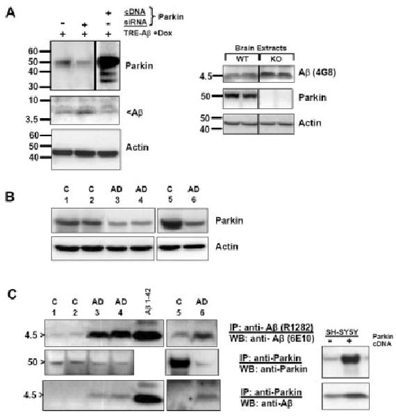
The significance of intracellular beta-amyloid (Abeta(42)) accumulation is increasingly recognized in Alzheimer's disease (AD) pathogenesis. Abeta removal mechanisms that have attracted attention include IDE/neprilysin degradation and antibody-mediated uptake by immune cells. However, the role of the ubiquitin-proteasome system (UPS) in the disposal of cellular Abeta has not been fully explored. The E3 ubiquitin ligase Parkin targets several proteins for UPS degradation, and Parkin mutations are the major cause of autosomal recessive Parkinson's disease. We tested whether Parkin has cross-function to target misfolded proteins in AD for proteasome-dependent clearance in SH-SY5Y and primary neuronal cells. Wild-type Parkin greatly decreased steady-state levels of intracellular Abeta(42), an action abrogated by proteasome inhibitors. Intracellular Abeta(42) accumulation decreased cell viability and proteasome activity. Accordingly, Parkin reversed both effects. Changes in mitochondrial ATP production from Abeta or Parkin did not account for their effects on the proteasome. Parkin knock-down led to accumulation of Abeta. In AD brain, Parkin was found to interact with Abeta and its levels were reduced. Thus, Parkin is cytoprotective, partially by increasing the removal of cellular Abeta through a proteasome-dependent pathway.


Figures

Fig. 1
Fig. 1
AI: Parkin expression systems. A: Schematic representation of the lentiviral construct encoding WT and mutant (T240R and ΔUbl) myc-tagged Parkin. B: Western blot of whole-cell extracts demonstrating expression of lentiviral Parkin constructs WT, T240R, and ΔUbl. Immunofluorescence analysis of SH-SY5Y cells transduced for Parkin (D) (rhodamine fluorescence) and its accompanying myc tag (C) (FITC fluorescence) at ×40. F: Hoechst 33258. G: Hoffman modulation contrast. H: Anti-Parkin labeling reveals expression in both the cytosol and the nucleus of SH-SY5Y cells (confocal 0.1 μm optical section). I: Subcellular fractionation of Adv-Aβ42-infected SH-SY5Y cells to localize β-amyloid and Parkin expression. Equal amounts of protein from each fraction were resolved on 4–12% Bis-Tris NuPAGE gels, so the levels shown would not necessarily reflect their relative absolute contributions to total cellular protein. Endogenous Parkin is found in all compartments, especially the mitochondrial fraction (P2), as characterized by cytochrome C localization. Heterologous Aβ42 is similarly abundant in mitochondria but also resides with Parkin in membrane-associated fractions (P3). Aβ and Parkin levels found in the crude nuclear fraction (P1) likely represent a contribution from either unbroken cells and/or nuclear membrane breakdown, as per cytochrome C contamination.
Fig. 2
Fig. 2
42 and Parkin levels in SH-SY5Y cells and primary cortical neurons in the presence of proteasome inhibitors. A: Cellular expression of β-amyloid under the inducible control of an adenovirus-based vector system shows equal partitioning into detergent (TX-100)-soluble and -insoluble fractions. SH-SY5Y cells infected at 100 m.o.i. and induced with 1 μg/ml doxycycline for 24 hr. The pellet was solubilized in 25% formic acid. B: Western blot analysis showing the effects of Parkin on steady-state levels of soluble (S) and insoluble (I) Aβ42 in SH-SY5Y cells. Aβ42 levels (top gel) detected with 6E10 antibody and Parkin levels (bottom gel) after stripping and reprobing with anti-Parkin antibody (PRK8). Wild-type but not mutant Parkin greatly reduces levels of Aβ42 in both compartments. Equal amounts of each soluble fraction sample (lanes 2, 4, 6, 8) were loaded, as shown in the actin blot below. C: Proteasome inhibition with MG132 (M) or lactacystin (L) increases steady-state levels of Aβ42. D: Parkin expression mitigates whole-cell Aβ42 levels, except where MG132 is present. E: Densitometric analysis of Parkin activity to lower Aβ levels and abrogation by inhibition of the proteasome (P < 0.02 compared with Parkin alone, n = 2 experiments). F, G: Parkin expression decreases steady-state levels of Aβ42 in whole-cell soluble (S) and insoluble (I) fractions prepared from primary neuronal cultures. An equal amount of protein (30 μg) corresponding to soluble fractions was loaded into each well on a 4–12% NuPAGE Bis-Tris gel. The insoluble fraction contained the entire cell pellet. Note that proteasome inhibition does not affect Parkin levels.
Fig. 3
Fig. 3
Cell death in Aβ42-stressed neuroblastoma cells: rescue by WT Parkin. A: MTT reduction is inhibited in Aβ-expressing SH-SY5Y cells in the presence of doxycycline (bar 3) and reversed only by WT Parkin (bar 5) and not its mutant forms. No reversal is obtained in the presence of MG132 (bar 10). Mean ± SD, n = 5, P < 0.05, Mann-Whitney test. *Significantly different from control (bar 1), #significantly different from Aβ42-stressed cells (bar 3). All values expressed as percentage of control. B: Synthetic Aβ, when added to SH-SY5Y cell medium (10 μM), is equally toxic but not rescued through Parkin expression; n = 4. C: In the absence of serum components, Parkin similarly reduces levels of intracellular Aβ. The lower amount of cellular Aβ under serum-free conditions likely reflects both stress and reduced uptake of exogenous Aβ present in the serum.
Fig. 4
Fig. 4
42 impairs 20S proteasome activity while associating with it and is rescued by Parkin. A: The activity of the 20S proteasome is inhibited in Aβ42-stressed SH-SY5Y cells (bar 5 vs. bar 4, *P < 0.05) and rescued by WT Parkin (bar 7 vs. bar 5, #P < 0.05) but not up to stimulated levels afforded by Parkin alone (bar 10 vs. bar 4, *P < 0.05). WT Parkin cannot overcome MG132 or lactacystin toxicities, under conditions with or without Aβ42 (bars 11 and 14 vs. 2, 3, and 6). Triplicate of n = 2 experiments. B: Aβ association with the proteasome. SH-SY5Y cells were induced to express β-amyloid, and the 20S proteasome was immunoprecipitated with an antibody recognizing multiple α and β subunits in the 27–30-kD range. The IPs were solubilized in sample buffer lacking (−) or containing (+) the reducing agent β-mercaptoethanol (β-ME). More Aβ immuno-reactivity is released from association with the anti-20S precipitate in the presence of β-ME (lane 6 vs. 5). C: Left: Effects of Parkin on simultaneous Aβ-affected and mitochondria-/proteasome-inhibited cells. The mitochondrial inhibitors rotenone and CCCP do not affect chymotrypsin-like proteasome activity, in contradistinction to Aβ. However, in their presence, Parkin stimulation is reduced. [Mean ± 1 SD, n = 5: vs. ctrl (bar 1) *P < 0.05, vs. Aβ alone (bar 4), #P < 0.05, Mann-Whitney test.] Note that bars 1, 2, 3, 4 correspond to bars 1, 2, 10, 5 in A. Right: Parkin reverses ATP loss in Aβ42- and mitochondrial poison-stressed cells (bars 3, 4 and bars 5–8, respectively). [Mean ± 1SD, n = 3, vs. Aβ, rotenone, or CCCP alone *P < 0.05, Mann-Whitney test.]
Fig. 5
Fig. 5
Parkin accelerates Aβ degradation. A: Left: Knock-down of Parkin in cell culture increases accumulation of cellular Aβ. SH-SY5Y cells were transfected with 80 pM Parkin siRNA for 48 hr simultaneously with inducible Aβ cDNA transfection. A companion transfection was performed with Parkin cDNA (a shorter exposure for this lane is shown). Duplicate experiments were performed with identical results. Right: Aβ expression in Parkin knockout mice. Whole-brain lysate from a pair of 6–7-month-old Parkin null mice and control littermates were immunoprecipitated with anti-Aβ (4G8) and immunoblotted with an anti-rodent Aβ-specific antibody (upper panel). Parkin immunoblot is shown below. A younger pair of mice gave similar results (not shown). B: Human autopsy brain samples from sporadic AD patients are deficient in Parkin. Lysates (20 μg protein) were fractionated by Western and probed for Parkin (top) or actin (loading control, bottom). Cortical samples corresponding to control cases 1 and 2 and AD cases 3 and 4 were from frontal pole (BA 10). In an additional experiment, a different control (case 5) and AD (case 6) sample were obtained from inferomedial temporal cortex (BA 20/36). All AD cases were Braak stage V. C: Immunoprecipitates (IP) of Aβ (top) or Parkin (middle and bottom) were prepared from lysates (200 μg protein) corresponding to the same cases as in B. The lower levels of Parkin were nonetheless associated with larger quantities of Aβ in all AD cases compared with control (bottom panel). Inset: Parkin was immunoprecipitated from SH-SY5Y extracts prepared from cells transfected with either control (−) or Parkin (+) vector and containing Aβ42. Western blots were probed for either Parkin (top) or β-amyloid (bottom). Increased Parkin leads to increased coimmunoprecipitable Aβ. Forty micrograms of total protein; higher exposure reveals oligomers in the pull down (results not shown).

Similar articles

Cited by

References

    1. Ardley HC, Scott GB, Rose SA, Tan NG, Markham AF, Robinson PA. Inhibition of proteasomal activity causes inclusion formation in neuronal and nonneuronal cells overexpressing Parkin. Mol Biol Cell. 2003;14:4541–4556. - PMC - PubMed
    1. Bonifacino JS, Weissman AM. Ubiquitin and the control of protein fate in the secretory and endocytic pathways. Annu Rev Cell Dev Biol. 1998;14:19–57. - PMC - PubMed
    1. Caspersen C, Wang N, Yao J, Sosunov A, Chen X, Lustbader JW, Xu HW, Stern D, McKhann G, Yan SD. Mitochondrial Abeta: a potential focal point for neuronal metabolic dysfunction in Alzheimer's disease. FASEB J. 2005;19:2040–2041. - PubMed
    1. Chafekar SM, Zwart R, Veerhuis R, Vanderstichele H, Baas F, Scheper W. Increased Abeta1–42 production sensitizes neuroblastoma cells for ER stress toxicity. Curr Alzheimer Res. 2008;5:469–474. - PubMed
    1. Chung KK, Zhang Y, Lim KL, Tanaka Y, Huang H, Gao J, Ross CA, Dawson VL, Dawson TM. Parkin ubiquitinates the alpha-synuclein-interacting protein, synphilin-1: implications for Lewy-body formation in Parkinson disease. Nat Med. 2001;7:1144–1150. - PubMed

Publication types

MeSH terms