. 2010 Jan;88(1):86-94.
doi: 10.1002/jnr.22179.

Postnatal development of NT3 and TrkC in mouse ventral cochlear nucleus

Affiliations

Postnatal development of NT3 and TrkC in mouse ventral cochlear nucleus

J Feng et al. J Neurosci Res. 2010 Jan.

Abstract

In the developing nervous system, neurotrophin 3 (NT3) and brain-derived neurotrophic factor (BDNF) have been shown to interact with each other and with different parts of a neuron or glia and over considerable distances in time and space. The auditory system provides a useful model for analyzing these events, insofar as it is subdivided into well-defined groups of specific neuronal types that are readily related to each other at each stage of development. Previous work in our laboratory suggested that NT3 and its receptor TrkC in the mouse cochlear nucleus (CN) may be involved in directing neuronal migration and initial targeting of inputs from cochlear nerve axons in the embryo. NT3 is hard to detect soon after birth, but TrkC lingers longer. Here we found NT3 and TrkC around P8 and the peak around P30. Prominent in ventral CN, associated with globular bushy cells and stellate cells, they were localized to different subcellular sites. The TrkC immunostain was cytoplasmic, and that of NT3 was axonal and perisomatic. TrkC may be made by CN neurons, whereas NT3 has a cochlear origin. The temporal pattern of their development and the likelihood of activity-dependent release of NT3 from cochlear axons suggest that it may not be critical in early synaptogenesis; it may provide long-term trophic effects, including stabilization of synapses once established. Activity-related regulation could coordinate the supply of NT3 with inner ear activity. This may require interaction with other neurotrophins, such as BDNF.


Figures

Fig. 1
Fig. 1
Left: TrkC staining of a transverse section through the middle one-third of CN in a montage. Staining is most intense in and around PV. Right: A comparable level of the mouse atlas identifies the different regions (Trettel and Morest, 2001). A, anterior part of posteroventral CN (PVCN); AD, anterodorsal part of PVCN; AS, acoustic striae; AV, anteroventral part of PVCN; C, cerebellum; CN, cochlear nucleus; F, fusiform layer of DCN; M, molecular layer of dorsal (D) CN; P, polymorphic layer of DCN; PV, ventral part of posterior anteroventral CN (AVCN); S, small cell shell; TC, taenia choroidea. Scale bars = 200 μm.
Fig. 2
Fig. 2
TrkC staining of PV. The perikaryal cytoplasm is stained at all ages, albeit to different degrees. Single arrows, globular bushy cells; double arrows, stellate cell; large arrows, cells in the insets. The P30 inset shows a typical globular bushy cell with unstained eccentric nucleus. The large arrowhead points to the nerve terminal region around the soma, which is unstained, next to a stained astrocyte (thin arrows point to the cell body and astrocytic processes). Next to the astrocyte are two oligodendrocytes (asterisks). The cytoplasmic stain in both P30 and P14 insets is granular, unlike the cell in P8 (lower inset). In the P8 main panel and the upper inset, astrocytic processes (thin arrow) between two neurons are stained. Scale bar = 20 μm; 5 μm for insets.
Fig. 3
Fig. 3
NT3 staining of PV. Perisomatic endings are stained at all ages. Same symbols for cell types as in Figure 2. Cochlear axons at P30 are heavily stained (thin arrow) along with clusters of nerve terminals at P30 and P8 and the incoming terminals at P14 (triangles in the insets). The arrowheads in P14 and P8 point to nerve terminals approaching their target sites. Scale bar = 20 μm; 5 μm for insets.
Fig. 4
Fig. 4
Fluoresecent double labeling for NT3 (green) and SV2 (red) to show colocalization (yellow) in the PV region (P60). Left arrow, axons abundant with NT3; right arrow, NT3 in the perisomatic region. SV2 labels nerve terminals (double arrows); many of them are also labeled for NT3 (arrowheads). Asterisk, example of a globular bushy cell. Scale bar = 20 μm.
Fig. 5
Fig. 5
Comparison of perisomatic staining of NT3 and perikaryal location of TrkC in globular bushy cells (thick arrows; P30). Images taken at high magnification (×63 oil). Astrocytes are also stained for TrkC (thin arrows). Inset: A cell with granular cytoplasm from the same section in another image. Scale bar = 20 μm.
Fig. 6
Fig. 6
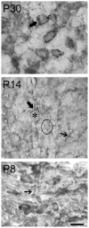
Synaptogenesis in PV as shown by SV2 staining. Single focal planes are shown from through-focus stacks of images (see Materials and Methods). At P30, the mature synaptic endings from the cochlear nerve, the endbulbs of Held, thickly enclose a typical globular bushy cell soma (thick arrow). The jagged line indicates the position of the eccentric nucleus, which could be visualized at another focal plane (compare with the inset in Fig. 2). At P14, an immature endbulb of Held (thick arrow) partially encloses a globular bushy cell (asterisk). Single processes of cochlear nerve fibers (thin arrow) and clusters of cochlear nerve fiber endings (circle) end freely in the neuropil, short of their targets. At P8, a single branching cochlear nerve fiber forms a cluster of endings in the neuropil (thin arrow). Scale bar = 20 μm.

References

    1. Agerman K, Hjerling-Leffler J, Blanchard MP, Scarfone E, Canlon B, Nosrat C, Ernfors P. BDNF gene replacement reveals multiple mechanisms for establishing neurotrophin specificity during sensory nervous system. Development. 2003;130:1479–1491. - PubMed
    1. Althaus HH, Richter-Landberg C. Glial cells as targets and producers of neurotrophins. Int Rev Cytol. 2000;197:203–277. - PubMed
    1. Bianchi LM, Conover JC, Fritzsch B, DeChiara T, Lindsay RM, Yancopoulos GD. Degeneration of vestibular neurons in late embryogenesis of both heterozygous and honoaygous BDNF null mutant mice. Development. 1996;122:1965–1973. - PubMed
    1. Brumwell CL, Hossain WA, Morest DK, Bernd P. Role for basic fibroblast growth factor (FGF-2) and TrkB expression in the development of the inner ear: in vitro and in situ studies. Exp Neurol. 1999;162:121–145. - PubMed
    1. Brumwell CL, Hossain WA, Morest DK, Wolf B. Biotinidase reveals the morphogenetic sequence in cochlea and cochlear nucleus of mice. Hear Res. 2005;209:104–121. - PubMed

Publication types