. 2009 Jun;36(6):2193-205.
doi: 10.1118/1.3116776.

Measurement of MRI scanner performance with the ADNI phantom

Affiliations

Measurement of MRI scanner performance with the ADNI phantom

Jeffrey L Gunter et al. Med Phys. 2009 Jun.

Abstract

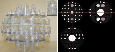
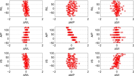
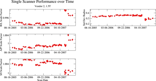
The objectives of this study are as follows: to describe practical implementation challenges of multisite, multivendor quantitative studies; to describe the MRI phantom and analysis software used in the Alzheimer's Disease Neuroimaging Initiative (ADNI) study, illustrate the utility of the system for measuring scanner performance, the ability to assess gradient field nonlinearity corrections: and to recover human brain images without geometric scaling errors in multisite studies. ADNI is a large multicenter study with each center having its own copy of the phantom. The design of the phantom and analysis software are presented as results from predistribution systematics studies and results from field experience with the phantom at 58 enrolling ADNI sites over a 3 year period. The estimated coefficients of variation intrinsic to measurements of geometry in a single phantom are in the range of 3-5 parts in 10(4). Phantom measurements accurately detect linear and nonlinear scaling in images. Gradient unwarping methods are readily assessed by phantom nonlinearity measurements. Phantom-based scaling correction reduces observed geometric drift in human images by one-third or more. Repair or replacement of phantoms between scans, however, is a confounding factor. The ADNI phantom can be used to assess both scanner performance and the validity of postprocessing image corrections in order to reduce systematic errors in human images. Reduced measurement errors should decrease measurement bias and increase statistical power for measurements of rates of change in the brain structure in AD treatment trials. Perhaps the greatest practical value of incorporating ADNI phantom measurements in a multisite study is to identify scanner errors through central monitoring. This approach has resulted in identification of system errors including sites misidentification of their own gradient hardware and the disabling of autoshim, and a miscalibrated laser alignment light. If undetected, these errors would have contributed to imprecision in quantitative metrics at over 25% of all enrolling ADNI sites.


Figures

Figure 1
Figure 1
ADNI phantom. A photograph of the internal components of the ADNI phantom is shown. Each of the spheres is filled with a copper sulfate solution. The colored spheres contain differing solution concentrations. The small inset provides a detailed view of a single sphere and postcomponent. A triplanar view of a phantom image acquired with the MP-RAGE used in the ADNI protocol is also shown.
Figure 2
Figure 2
Qualitative evaluation of geometric performance. Plots of sphere position (vertical axes) versus displacement (horizontal axes) in each cardinal direction provide qualitative image distortion information. All lengths are measured in mm, and the position origin is MR scanner isocenter.
Figure 3
Figure 3
Calibration exercise. Sets of plots with data before (left) and after (right) an exercise in scanner calibration are shown. After calibration, the dependence of position residuals (the horizontal axes in the subplots) on position (the vertical axes in the subplots) was greatly reduced. The two obvious outlier points in the AP versus ΔAP subplots were due to manufacturing defects and subsequently repaired.
Figure 4
Figure 4
Construction variability. Histograms of normalized phantom size for the initial 66 production phantoms used in the ADNI study are shown.
Figure 5
Figure 5
Nonlinearity estimates. The dependence of the residual radius distribution for different orders of polynomial displacement field is shown for a scan with 2D (left) and 3D (middle) gradient warping corrections. In these plots, each horizontal row contains a histogram of 160 residual radii for a deformation field of given polynomial order (which is indicated on the vertical axes). The sizes of the boxes in the plots are proportional to the density of points. The rightmost plot presents the standard deviation of the distributions for data with 2D warping correction (open stars) and for 3D warping correction (solid red circles).
Figure 6
Figure 6
Longitudinal tracking of individual scanner from vendor 2 with phantom measurements. The left panel demonstrates scale factors along each cardinal axis. The right panels show the standard deviation of residual radius (nonlinearity). The system was recalibrated in early and mid-2006 as well as mid-2007 when the system underwent an upgrade. After the upgrade, the standard deviation of residual radius metric for nonlinearity was decreased.
Figure 7
Figure 7
Longitudinal tracking of individual scanner from vendor 3 with phantom measurements. The left panel demonstrates scale factors along each cardinal axis. The right panels show the standard deviation of residual radius (nonlinearity). Prior to mid-2007, the protocol for this vendor was errantly distributed with autoshimming disabled, a fact reflected in the larger variation in the SI scale factors. Note that the vertical range for the SI scale factor time course is larger than for other dimensions.
Figure 8
Figure 8
Summary of scanner performance for more than 2200 phantom scans. A pooled-variance approach is used to estimate the stability of gradient performance factoring out discrete changes generally due to scanner recalibration. Symbols are plotted at the mean scale value over all values, and error bars indicate the square root of the pooled variance. System number is an arbitrary enumeration. RL calibration appears less consistent across scanners for vendor 1 than for other vendors. The SI per scanner error bars for vendor 3 are much larger than for other vendors and other directions. Scanners from vendor 2 are from two different models and the data are clustered by model in the SI mean scale factors.
Figure 9
Figure 9
Use of phantom measurements to correct within-scanner linear scaling changes in human images. Histograms of intrasubject coregistration scale factors from 1.5 T scanners with and without phantom-based voxel size adjustment are shown. The upper (lower) histograms are without (with) correction. Correction reduces the widths of the distributions. The vertical dashed lines are located at 1.00, the ideal intrasubject scale factor.
Figure 10
Figure 10
Use of phantom measurements to perform absolute scaling of human images across scanner. Histograms of intrasubject coregistration scale factors for image pairs with one scan acquired at 3 T and the other at 1.5 T are shown. The upper (lower) histograms are without (with) correction. Correction reduces the widths of the distributions.

References

    1. Baldwin L. N., Wachowicz K., Thomas S. D., Rivest R., and Fallone B. G., “Characterization, prediction, and correction of geometric distortion in 3 T MR images,” Med. Phys. MPHYA6 34(2), 388–399 (2007).10.1118/1.2402331 - DOI - PubMed
    1. Breeuwer M. M., Holden M., and Zylka W., “Detection and correction of geometric distortion in 3D MR images,” Proc. SPIE PSISDG 4322, 1120–1120 (2001).
    1. Chen C. C., Wan Y. L., Wai Y. Y., and Liu H. L., “Quality assurance of clinical MRI scanners using ACR MRI phantom: Preliminary results,” J. Digit Imaging ZZZZZZ 17(4), 279–284 (2004).10.1007/s10278-004-1023-5 - DOI - PMC - PubMed
    1. Colombo P., Baldassarri A., Del Corona M., Mascaro L., and Strocchi S., “Multicenter trial for the set-up of a MRI quality assurance programme,” Magn. Reson. Imaging MRIMDQ 22(1), 93–101 (2004).10.1016/j.mri.2003.04.001 - DOI - PubMed
    1. Firbank M. J., Harrison R. M., Williams E. D., Coulthard A., Britson P. J., Gunter J. L., and Ward C. P., “Quality assurance for MRI: Practical experience,” Br. J. Radiol. BJRAAP 73(868), 376–383 (2000). - PubMed

Publication types

MeSH terms