

Scatter correction for cone-beam CT in radiation therapy
- PMID: 19610315
- PMCID: PMC2832067
- DOI: 10.1118/1.3130047
Scatter correction for cone-beam CT in radiation therapy
Abstract
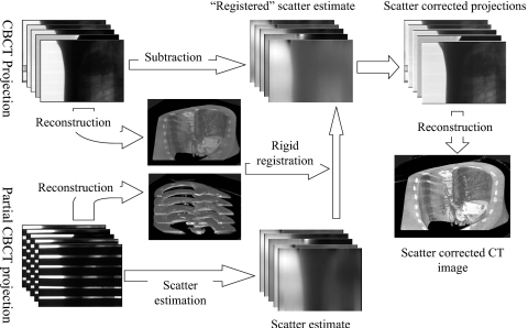
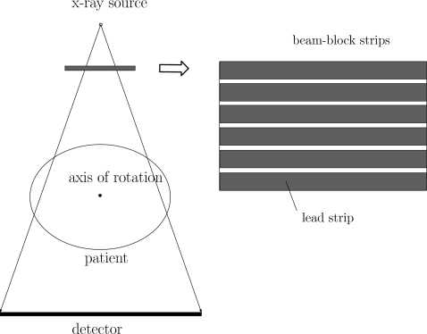
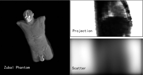
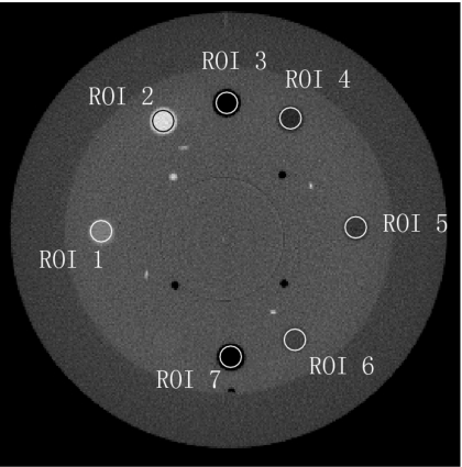
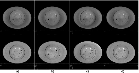
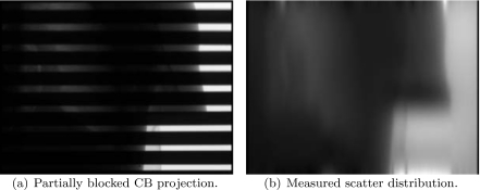
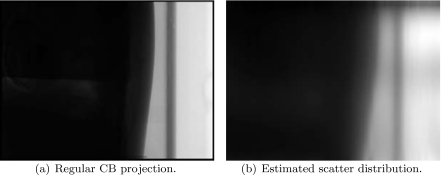
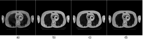
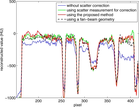
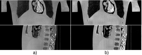
Cone-beam CT (CBCT) is being increasingly used in modern radiation therapy for patient setup and adaptive replanning. However, due to the large volume of x-ray illumination, scatter becomes a rather serious problem and is considered as one of the fundamental limitations of CBCT image quality. Many scatter correction algorithms have been proposed in literature, while a standard practical solution still remains elusive. In radiation therapy, the same patient is scanned repetitively during a course of treatment, a natural question to ask is whether one can obtain the scatter distribution on the first day of treatment and then use the data for scatter correction in the subsequent scans on different days. To realize this scatter removal scheme, two technical pieces must be in place: (i) A strategy to obtain the scatter distribution in on-board CBCT imaging and (ii) a method to spatially match a prior scatter distribution with the on-treatment CBCT projection data for scatter subtraction. In this work, simple solutions to the two problems are provided. A partially blocked CBCT is used to extract the scatter distribution. The x-ray beam blocker has a strip pattern, such that partial volume can still be accurately reconstructed and the whole-field scatter distribution can be estimated from the detected signals in the shadow regions using interpolation/extrapolation. In the subsequent scans, the patient transformation is determined using a rigid registration of the conventional CBCT and the prior partial CBCT. From the derived patient transformation, the measured scatter is then modified to adapt the new on-treatment patient geometry for scatter correction. The proposed method is evaluated using physical experiments on a clinical CBCT system. On the Catphan 600 phantom, the errors in Hounsfield unit (HU) in the selected regions of interest are reduced from about 350 to below 50 HU; on an anthropomorphic phantom, the error is reduced from 15.7% to 5.4%. The proposed method is attractive in applications where a high CBCT image quality is critical, for example, dose calculation in adaptive radiation therapy.
Figures

Similar articles
-
Shading correction for on-board cone-beam CT in radiation therapy using planning MDCT images.Med Phys. 2010 Oct;37(10):5395-406. doi: 10.1118/1.3483260. Med Phys. 2010. PMID: 21089775
-
Combining scatter reduction and correction to improve image quality in cone-beam computed tomography (CBCT).Med Phys. 2010 Nov;37(11):5634-44. doi: 10.1118/1.3497272. Med Phys. 2010. PMID: 21158275
-
Quantitative cone-beam CT imaging in radiation therapy using planning CT as a prior: first patient studies.Med Phys. 2012 Apr;39(4):1991-2000. doi: 10.1118/1.3693050. Med Phys. 2012. PMID: 22482620 Free PMC article.
-
A general framework and review of scatter correction methods in x-ray cone-beam computerized tomography. Part 1: Scatter compensation approaches.Med Phys. 2011 Jul;38(7):4296-311. doi: 10.1118/1.3599033. Med Phys. 2011. PMID: 21859031 Review.
-
Technical aspects of dental CBCT: state of the art.Dentomaxillofac Radiol. 2015;44(1):20140224. doi: 10.1259/dmfr.20140224. Dentomaxillofac Radiol. 2015. PMID: 25263643 Free PMC article. Review.
Cited by
-
Development and validation of a scatter-corrected CBCT image-guided method for cervical cancer brachytherapy.Front Oncol. 2022 Oct 27;12:942016. doi: 10.3389/fonc.2022.942016. eCollection 2022. Front Oncol. 2022. PMID: 36387118 Free PMC article.
-
Antiscatter grids in mobile C-arm cone-beam CT: effect on image quality and dose.Med Phys. 2012 Jan;39(1):153-9. doi: 10.1118/1.3666947. Med Phys. 2012. PMID: 22225284 Free PMC article.
-
Improving CBCT image quality to the CT level using RegGAN in esophageal cancer adaptive radiotherapy.Strahlenther Onkol. 2023 May;199(5):485-497. doi: 10.1007/s00066-022-02039-5. Epub 2023 Jan 23. Strahlenther Onkol. 2023. PMID: 36688953 Free PMC article.
-
John's Equation-based Consistency Condition and Corrupted Projection Restoration in Circular Trajectory Cone Beam CT.Sci Rep. 2017 Jul 7;7(1):4920. doi: 10.1038/s41598-017-05249-5. Sci Rep. 2017. PMID: 28687756 Free PMC article.
-
Comparison of Supervised and Unsupervised Approaches for the Generation of Synthetic CT from Cone-Beam CT.Diagnostics (Basel). 2021 Aug 9;11(8):1435. doi: 10.3390/diagnostics11081435. Diagnostics (Basel). 2021. PMID: 34441369 Free PMC article.
References
-
- Fox T. R., Nisius D. T., Aradate H., and Saito Y., “Practical x-ray scatter measurements for volume CT detector design,” in Medical Imaging 2001: Physics of Medical Imaging, edited by Antonuk Larry E. and Yaffe Martin J.
- [Proc. SPIE PSISDG 4320, 808–814 (2009)].
-
- Lee L., Le Q.-T., and Xing L., “Retrospective IMRT dose reconstruction based on cone-beam ct and mlc log-file,” Int. J. Radiat. Oncol., Biol., Phys. IOBPD3 70(2), 634–644 (2008). - PubMed
Publication types
MeSH terms
Grants and funding
LinkOut - more resources
Full Text Sources
Other Literature Sources
