Review
. 2009 Oct 16;4(10):811-27.
doi: 10.1021/cb900156h.

Conformational dynamics in the Acyl-CoA synthetases, adenylation domains of non-ribosomal peptide synthetases, and firefly luciferase

Affiliations
Review

Conformational dynamics in the Acyl-CoA synthetases, adenylation domains of non-ribosomal peptide synthetases, and firefly luciferase

Andrew M Gulick. ACS Chem Biol. .

Abstract

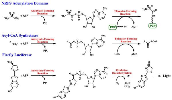
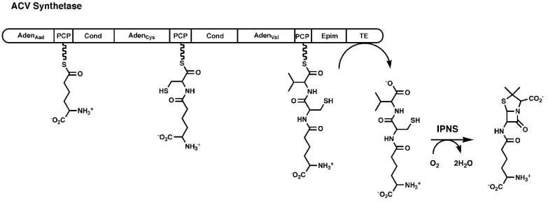
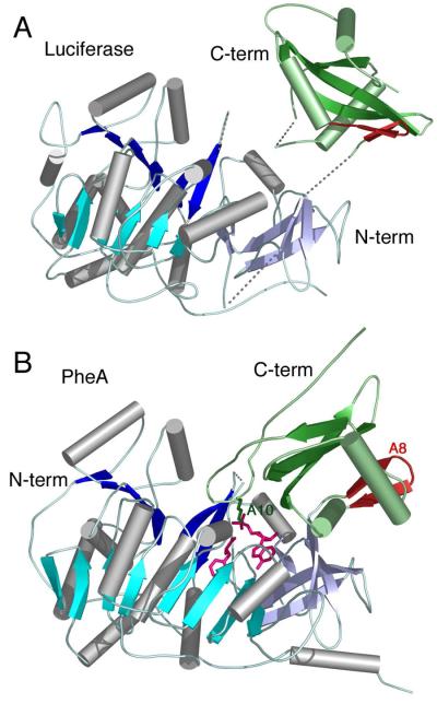
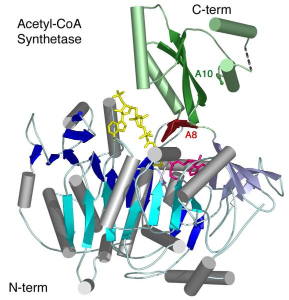
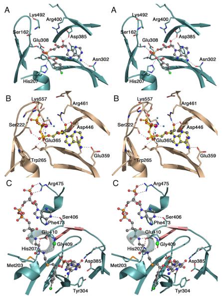
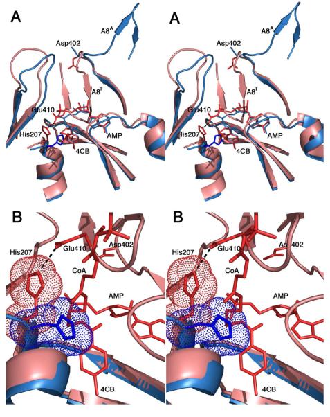
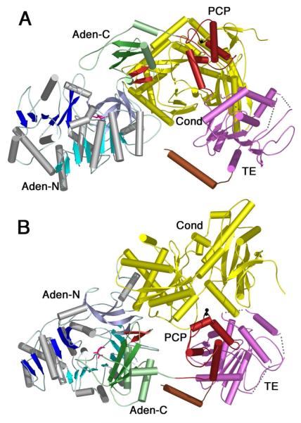
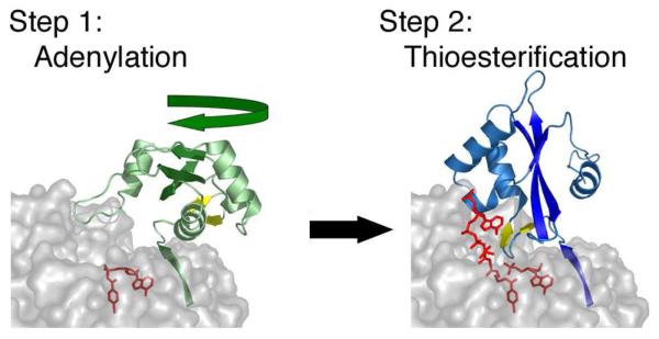
The ANL superfamily of adenylating enzymes contains acyl- and aryl-CoA synthetases, firefly luciferase, and the adenylation domains of the modular non-ribosomal peptide synthetases (NRPSs). Members of this family catalyze two partial reactions: the initial adenylation of a carboxylate to form an acyl-AMP intermediate, followed by a second partial reaction, most commonly the formation of a thioester. Recent biochemical and structural evidence has been presented that supports the use by this enzyme family of a remarkable catalytic strategy for the two catalytic steps. The enzymes use a 140 degrees domain rotation to present opposing faces of the dynamic C-terminal domain to the active site for the different partial reactions. Support for this domain alternation strategy is presented along with an explanation of the advantage of this catalytic strategy for the reaction catalyzed by the ANL enzymes. Finally, the ramifications of this domain rotation in the catalytic cycle of the modular NRPS enzymes are discussed.


Figures

Figure 1
Figure 1. Reactions catalyzed by the ANL Superfamily of adenylating enzymes
Chemical reactions catalyzed by the three subfamilies of the ANL adenylating enzymes. All three reactions include an initial, adenylate-forming reaction to form an acyl-, amino acyl-, or aryl-adenylate with the release of inorganic pyrophosphate. The adenylate intermediate reacts in a second partial reaction to release AMP. For NRPS adenylation domains, the pantetheine cofactor bound to an NRPS peptidyl carrier domain is represented by the linker and SH group.
Figure 2
Figure 2. Modular Organization of the Non-Ribosomal Peptide Synthetases
A schematic representation is shown of the ACV synthetase, a three-module protein that is responsible for the synthesis of the linear tripeptide of α-aminoadipic acid (Aad), cysteine, and valine. The linear peptide is subsequently cyclized by the enzyme isopenicillin N synthase. Module 1 contains the adenylation domain and PCP for Aad. Module 2 contains the adenylation and PCP domain for cysteine, as well as the condensation domain that forms the peptide bond between Aad and Cys. The third module contains the adenylation and PCP domain for valine, an epimerization domain that converts l-Val to d-Val, and a condensation domain that transfers the upstream dipeptide to d-Val. The protein terminates with a thioesterase domain that releases the tripeptide.
Figure 3
Figure 3. Crystal Structures of A. Firefly Luciferase and B. PheA adenylation domain
Structures of the first two members of the ANL superfamily of enzymes to be characterized by crystallography. The proteins are aligned on the basis of the N-terminal domains. The structure of luciferase (1LCI) displayed an open conformation with few interactions between the N- and C-terminal domains. Several disordered loops are indicated with dashed lines. PheA was co-crystallized in the presence of ATP and phenylalanine (1AMU), and revealed a molecule of AMP and Phe in the active site (pink). The A8 loop, that follows the hinge for domain alternation is shown in red while the A10 lysine that is conserved throughout the entire family is shown in green. In both panels, the N-terminal domain is represented with β-sheets of different shades of blue, while the C-terminal domain is shown in green.
Figure 4
Figure 4. Crystallographic Structure of Acetyl-CoA synthetase
The structure is shown of bacterial acetyl-CoA synthetase (1PG4). The enzyme is oriented as other family members are in Figure 3 and includes the ligands adenosine-5′-propyl phosphate (pink) and CoA (yellow). The CoA nucleotide binds on the surface of the N-terminal domain while the pantetheine passes into the enzyme active site via the pantetheine tunnel. The A8 loop that follows the hinge is shown in red. The Cα and Cβ positions of Lys609, the A10 lysine, are shown in green. The N- and C-terminal domains are colored as in Figure 3.
Figure 5
Figure 5. Active site and binding interactions of the ANL enzymes
A. Aryl-AMP binding site of 4CBL determined in the adenylate-forming conformation (3CW8). Conserved residues that interact with the adenylate are shown. Also interacting with the α-phosphate are Thr161, from the P-loop, and Thr307. Tyr304, behind the adenine ring is unlabeled. The side chain of the A10 lysine, Lys492, was disordered beyond Cβ. B. ATP binding site of medium chain Acyl-CoA synthetase in the adenylate-forming conformation (3C5E). The interaction of the Gly- and Ser/Thr-rich P-loop with the triphosphate of ATP is clearly demonstrated. The Mg2+ ion, which bridges the b- and g-phosphates, is shown in purple. Pairs of homologous residues between mAcs and 4CBL are (mAcs listed first with 4CBL residue in parentheses): Ser222 (Thr161), Thr223 (Ser162), Trp265 (His207), Glu359 (Asn302), Tyr361 (Tyr304), Thr364 (Thr307), Glu365 (Glu308), Asp446 (Asp385), and Arg461 (Arg400). C. Binding interactions between CoA and 4CBL observed in the thioester-forming conformation (3CW9). The CoA nucleotide binds on the surface of the 4CBL enzyme, with the nucleotide ring stacking against Phe473. The pantetheine passes to the interior of the protein where the CoA thiol can attack the adenylate intermediate. Here, the thioester bond is modeled by the thioether linkage of 4-chlorophenacyl-CoA. The two strands that form the A8 loop are shown in pink and the Cα position of the universally conserved glycine at position 409 is shown with a green sphere. The β-sheet like interaction between Gly408-Gly409 and the β-alanine moiety of the pantetheine group is indicated with dashed lines. The interaction between His207 and Glu410 that stabilizes the A4 aromatic group (His207) in the new side chain position is shown with a red dashed line. The first turn of the helix at 251-265 is depicted transparently to allow the pantetheine chain to be seen.
Figure 6
Figure 6. Impact of domain alternation on the orientation of the A4 aromatic residue
A. Superposition of the crystal structures of 4CBL in both the adenylate-forming (3CW8, blue) and thioester-forming (3CW9, pink) conformations. The view is oriented so that the reader is looking down the pantetheine tunnel into the active site. The A8 loop is shown in both conformations, indicated by the superscript (A8A and A8T for the adenylate- and thioester-forming conformations, respectively). The 4-chlorobenzoyl adenylate (blue) and the 4-chlorophenacyl thioester and AMP (red) are shown. The terminal β-alanine and cysteamine groups of the pantetheine chain are shown. In the thioester-forming conformation, the A8 loop rotates into the active site (A8T), where Glu410 interacts with His207 to rotate the side chain away from the 4CB molecule. B. Closer view of the active site. The orientation is rotated slightly from panel A allowing observation of more of the pantetheine chain. The His207 side chain is shown with van der Waals surface. In the adenylate-forming structure, His207 occludes the pantetheine group from approaching the active site.
Figure 7
Figure 7. Crystal structure of the SrfA-C termination module from Surfactin NRPS cluster
The structure contains the domains organized as Condensation-Adenylation-PCP-Thioesterase, from N- to C-terminus (2VSQ). The N-terminal domain is colored as other members of the ANL family with N-terminal domain containing β-sheets of blue and the C-terminal domain shown in green. The A8 loop is shown as the two-stranded β-sheet in red and a molecule of leucine is shown in pink in the adenylate-binding pocket. The Condensation domain (yellow), PCP domain (red) and thioesterase domain (purple) are shown. The C-terminal purification tag formed a helix that is shown in brown. The cofactor binding site, Ser1003, was mutated to an alanine and is shown in black. In panel A, the adenylation domain is oriented as for other members of the ANL family in Figures 3 and 4. The C-terminal domain most closely represents the adenylate-forming conformation, although it is opened by ~40° compared to other enzymes. In panel B, the image is rotated by ~90° around the X-axis to depict the presentation of the cofactor binding site to the condensation domain active site cleft. The pantetheine cofactor is not present in the structure yet would be unable to reach the adenylation domain active site without a large conformational change.

References

    1. McElroy WD, DeLuca M, Travis J. Molecular uniformity in biological catalyses. The enzymes concerned with firefly luciferin, amino acid, and fatty acid utilization are compared. Science. 1967;157:150–160. - PubMed
    1. Bar-Tana J, Rose G. Studies on medium-chain fatty acyl-coenzyme a synthetase. Enzyme fraction I: mechanism of reaction and allosteric properties. Biochem J. 1968;109:275–282. - PMC - PubMed
    1. Lipmann F. Enzymatic synthesis of acetyl phosphate. J Biol Chem. 1944;155:55–70.
    1. Berg P. Acyl adenylates; an enzymatic mechanism of acetate activation. J Biol Chem. 1956;222:991–1013. - PubMed
    1. Farrar WW, Plowman KM. Kinetics of acetyl-CA synthetase–1. Mode of addition of substrates. International J. Biochem. 1975;6:537–542.

Publication types