. 2009 Sep 14;423(1):41-52.
doi: 10.1042/BJ20090786.

Development of Dictyostelium discoideum is associated with alteration of fucosylated N-glycan structures

Affiliations

Development of Dictyostelium discoideum is associated with alteration of fucosylated N-glycan structures

Birgit Schiller et al. Biochem J. .

Abstract

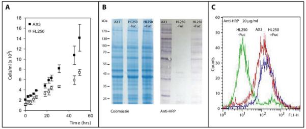
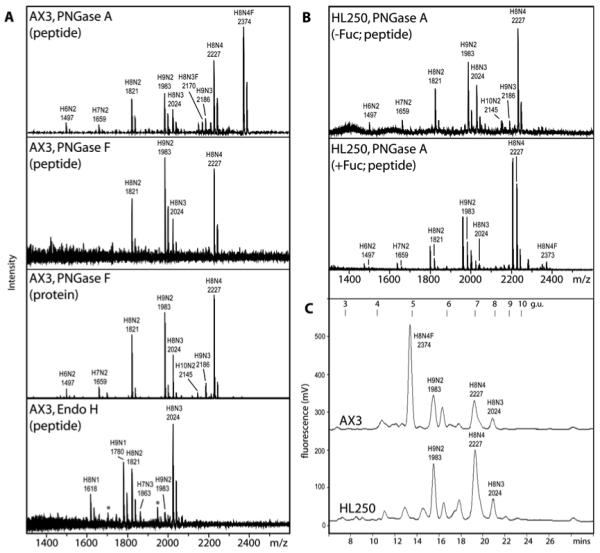
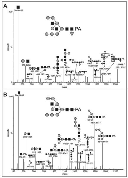
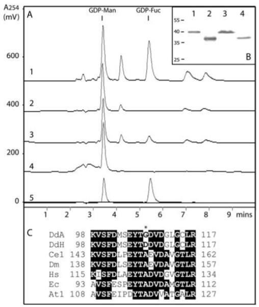
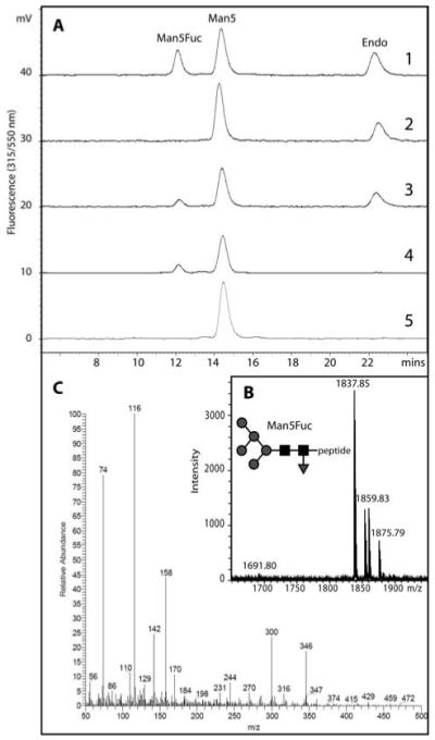
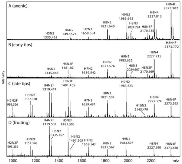
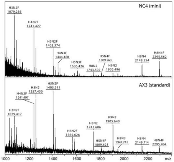
The social amoeba Dictyostelium discoideum has become established as a simple model for the examination of cell-cell interactions, and early studies suggested that shifts in glycosylation profiles take place during its life cycle. In the present study, we have applied HPLC and mass spectrometric methods to show that the major N-glycans in axenic cultures of the AX3 strain are oligomannosidic forms, most of which carry core fucose and/or intersecting and bisecting N-acetylglucosamine residues, including the major structure with the composition Man8GlcNAc4Fuc1. The postulated alpha1,3-linkage of the core fucose correlates with the cross-reactivity of Dictyostelium glycoproteins with a horseradish peroxidase antiserum; a corresponding core alpha1,3-fucosyltransferase activity capable of modifying oligomannosidic N-glycans was detected in axenic Dictyostelium extracts. The presence of fucose on the N-glycans and the reactivity to the antiserum, but not the fucosyltransferase activity, are abolished in the fucose-deficient HL250 strain. In later stages of development, N-glycans at the mound and culmination stages show a reduction in both the size and the degree of modification by intersecting/bisecting residues compared with mid-exponential phase cultures, consistent with the hypothesis that glycosidase and glycosyltransferase expression levels are altered during the slime mould life cycle.


Figures

Figure 1
Figure 1. Anti-horseradish peroxidase epitopes of axenically-cultured Dicytostelium discoideum
(A) The AX3 strain exhibits somewhat quicker growth during the pre-logarithmic phase of axenic culture in comparison to the HL250 (modC) strain; (B) Western blotting shows the presence of anti-HRP epitopes in axenically-grown AX3, but not in axenically-grown HL250, consistent with a defect in fucose metabolism in that strain; a partial recovery of anti-HRP epitopes was observed when HL250 cells were grown in the presence of exogenous fucose. The Coomassie-stained gel shows that protein loading was approximately equal. (C) Flow cytometry also shows the absence of anti-HRP epitopes in the HL250 strain grown in the absence of fucose (green) as compared to AX3 (red) or HL250 grown in the presence of exogenous fucose (blue).
Figure 2
Figure 2. The N-glycome of axenically-cultured Dicytostelium discoideum
(A) MALDI-TOF MS of pyridylaminated N-glycans from axenically-grown mid-log phase AX3 either released from glycopeptides using PNGase A, PNGase F or EndoH or from intact proteins treated with PNGase F showing the absence of fucosylated glycans from the latter three glycan pools. The peaks marked with an asterisk in the spectrum of the EndoH-released glycans are non-pyridylaminated forms of the Hex9HexNAc1 (m/z 1703) and Hex8HexNAc3 (m/z 1947) species. The data with pyridylaminated PNGase A-released glycans is compatible to those of the unlabelled, free N-glycans (see ESI-MS data in Supplementary Figure 2). (B) MALDI-TOF MS of PNGase A-released pyridylaminated N-glycans from axenically-grown mid-log phase HL250 grown in the absence or presence of exogenously-added fucose, showing the appearance of a fucosylated H8N4F glycan (m/z 2373) in the latter. C) RP-HPLC pyridylaminated PNGase A-released N-glycans from axenically-grown mid-log phase AX3 and HL250 grown in the absence of exogenous fucose shows a difference in elution properties; peaks were collected and subject to MALDI-TOF MS (major species are annotated) and compared to an isomaltooligosaccharide standard (g.u., glucose units). The major [M+Na]+ species are annotated in the form HxNyF0-1, where H signifies hexose, N is N-acetylhexosamine and F is fucose.
Figure 3
Figure 3. MS/MS analysis of the major N-glycans of axenically-grown mid-log cells
The major AX3 pyridylaminated N-glycans were purified by sequential NP-HPLC (Palpak) and RP-HPLC prior to analysis by LC-ESI-MS/MS; the MaxEnt3 transformed data for (A) the m/z 1176.93 [M+2H]2+ (Man8GlcNAc4Fuc1) and (B) the m/z 1103.41 [M+2H]2+ (Man8GlcNAc4) molecular ions are shown. Fragments of the postulated complete glycan structures are depicted using the nomenclature of the Consortium for Functional Glycomics (black square, N- acetylglucosamine; grey circle, mannose; grey triangle, fucose). The composition and linkages of the intact glycans were proven by GLC-MS (see Table 1); up to three mannose and one GlcNAc residues could be released by combined α-mannosidase and β-N-acetylhexosaminidase digestion (data not shown). Based on the expected prior processing by ER mannosidase, the Man8B isomer is taken as the basis for these structures.
Figure 4
Figure 4. Reconstitution of Dictyostelium GDP-Fucose biosynthesis in vitro
(A) Recombinant forms of the Dictyostelium GMD and GER encoded by cDNAs derived from either the AX3 or the HL250 strain were assayed in the following combinations: 1, AX3 GMD with AX3 GER; 2, HL250 GMD with AX3 GER; 3, AX3 GMD with HL250 GER; 4, HL250 GMD with HL250 GER; 5, overlaid chromatograms of GDP-Man and GDP-Fuc standards; other peaks in the RP-HPLC chromatograms derive from the bacterial lysates. (B) Expression of protein from all four clones was demonstrated by anti-His Western blotting: 1, AX3 GMD; 2, AX3 GER; 3, HL250 GMD; 4, HL250 GER. (C) Comparison of the protein sequences of various GDP-Man dehydratases in the region surrounding the site corresponding to the mutation in the HL250 GMD cDNA; DdA, Dicytostelium discoideum AX3 (residues 98-117); DdH, Dicytostelium discoideum HL250; Ce1, Caenorhabditis elegans GMD-1 (BRE-1); Dm, Drosophila melanogaster GMD; Hs, Homo sapiens GMD; Ec, Escherichia coli GMD; At1, Arabidopsis thaliana GMD1. Amino acids identical in a majority of the selected sequences are highlighted and the Gly → Asp change in the HL250 sequence is marked with an asterisk. On the other hand, the AX3 and HL250 ger cDNA sequences were found to be identical, although expression and activity levels differed in the experiments shown in panels A and B.
Figure 5
Figure 5. Demonstration of a novel core α1,3-fucosyltransferase activity in Dictyostelium
(A) A dansylated glycopeptide (dansyl-Asn-Ser-Man5GlcNAc2; Man5) was incubated in the presence of (1) a lysate of mid-log phase Dictyostelium AX3 cells and GDP-Fuc, (2) an AX3 lysate without GDP-Fuc, (3) an HL250 lysate and GDP-Fuc, (4) purified recombinant Caenorhabditis elegans FUT-1 and GDP-Fuc or (5) no enzyme. The shift to a lower retention time results in formation of a fucosylated glycopeptide (Man5Fuc). (B) The collected Man5Fuc fraction was subject to MALDI-TOF MS verifying the addition of fucose (m/z [M+Na]+ = 1837; as compared to the non-fucosylated from with m/z 1691). (C) The core 1,3-linkage of the transferred fucose was demonstrated by the presence of 3,4-disubstituted GlcNAc (2-deoxy-2-(N-methyl)acetamido-6-O-methyl-glucitol) in the obtained EI spectrum.
Figure 6
Figure 6. MALDI-TOF MS analysis of Dictyostelium N-glycans expressed during development
Pyridylaminated N-glycans derived from PNGase A digestion of axenic (A; 1.5 × 10⁶ cells per ml), early tips (B; 10-12 hrs), late tips (C; 16 hrs) and fruiting bodies (D; 24 hrs) of the AX3 strain were analysed by MALDI-TOF MS. Glycans from each stage were prepared two or three times independently; the relative intensities of these typical spectra were calculated and are presented in Supplementary Table 1. The major [M+Na]+ species are annotated in the form HxNyF0-1
Figure 7
Figure 7. Analysis of a putatively core α1,3-fucosylated N-glycan from Dictyostelium late tips
A) A normal-phase HPLC fraction (Palpak Type N; 8 g.u.) of late tip glycans released by PNGase A was analysed by RP-HPLC and shown to contain a major species with a retention time of 5.1 g.u., consistent with core α1,3-fucosylation. B) MALDI-TOF MS of the purified 5.1 g.u. fraction showed it to contain a species with m/z 1459.75 (Hex5HexNAc2Fuc1-PA; H+-adduct) as well as the corresponding Na+ and K+ adducts. C) The MS/MS fragmentation of this glycan shows the presence of an m/z 445 fragment consistent with the composition GlcNAc1Fuc1PA; key fragment ions are annotated. A comparable MS/MS spectrum was obtained from the corresponding glycan in a TSKgel Amide-80 (7.1 g.u.) fraction; the Hex5HexNAc2Fuc1 glycan was insensitive to both Aspergillus α1,2-mannosidase and bovine kidney α-fucosidase, but was sensitive to jack bean α-mannosidase (data not shown). The predicted structure of the glycan is shown according to the nomenclature of the Consortium for Functional Glycomics (black square, N-acetylglucosamine; grey circle, mannose; grey triangle, fucose).
Figure 8
Figure 8. Comparison of N-glycans from the fruiting bodies of axenic and non-axenic strains
Underivatised PNGase A-released N-glycans derived from NC4 and AX3 fruiting bodies were subject to MALDI-TOF MS in the positive mode. The NC4 glycans were prepared using a small-scale method as described, whereas the AX3 glycans are from the standard large-scale preparation (for the results with the corresponding pyridylaminated glycans, see Figure 7 and Supplementary Figure 4). The major [M+Na]+ species are annotated in the form HxNyF0-1.

Similar articles

Cited by

References

    1. Devreotes P. Dictyostelium discoideum: a model system for cell-cell interactions in development. Science. 1989;245:1054–1058. - PubMed
    1. Ziska SE, Henderson EJ. Cell surface oligosaccharides participate in cohesion during aggregation of Dictyostelium discoideum. Proc Natl Acad Sci U S A. 1988;85:817–821. - PMC - PubMed
    1. Frazier WA, Rosen SD, Reitherman RW, Barondes SH. Purification and comparison of two developmentally regulated lectins from Dictyostelium discoideum. Discoidin I and II. J Biol Chem. 1975;250:7714–7721. - PubMed
    1. Ivatt RL, Das OP, Henderson EJ, Robbins PW. Glycoprotein biosynthesis in Dictyostelium discoideum: developmental regulation of the protein-linked glycans. Cell. 1984;38:561–567. - PubMed
    1. Tschursin E, Riley GR, Henderson EJ. Differential regulation of glycoprotein sulfation and fucosylation during growth of Dictyostelium discoideum. Differentiation. 1989;40:1–9. - PubMed

Publication types