

Review
doi: 10.1042/BST0370707.
Electrodes modified with lipid membranes to study quinone oxidoreductases
Affiliations
- PMID: 19614580
- PMCID: PMC3827736
- DOI: 10.1042/BST0370707
Item in Clipboard
Review
Electrodes modified with lipid membranes to study quinone oxidoreductases
Biochem Soc Trans.
2009 Aug.
Abstract
Quinone oxidoreductases are a class of membrane enzymes that catalyse the oxidation or reduction of membrane-bound quinols/quinones. The conversion of quinone/quinol by these enzymes is difficult to study because of the hydrophobic nature of the enzymes and their substrates. We describe some biochemical properties of quinones and quinone oxidoreductases and then look in more detail at two model membranes that can be used to study quinone oxidoreductases in a native-like membrane environment with their native lipophilic quinone substrates. The results obtained with these model membranes are compared with classical enzyme assays that use water-soluble quinone analogues.
Figures
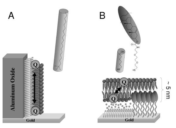
Model membrane systems used to study quinone oxidoreductases. (A) A hybrid bilayer on alumin oxide modified with trichloro(octadecyl)silane (OTS). This system has been used to study a peripheral membrane enzyme, pyruvate-ubiquinone oxidoreductase[32]. (B) A tethered bilayer lipid membrane (tBLM) formed on a mixed self-assembled monolayer of EO3-Cholesterol and 6-mercaptohexanol. This system has been used to study a ubiquinol oxidase, cytochrome bo3[33, 34].
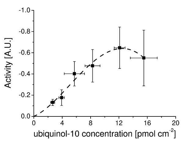
The enzyme activity of cytochrome bo3 from E. coli as a function of ubqiuinol-10 concentration in the membrane. Data determined using a tethered bilayer lipid membrane system as explained in the text. The dotted line is guide for the eye to highlight possible cooperative behaviour as described in the text. Details of the experiment are described in [34].
Similar articles
-
Quinone reduction and redox cycling catalysed by purified rat liver dihydrodiol/3 alpha-hydroxysteroid dehydrogenase.Biochem Pharmacol. 1992 Jul 22;44(2):341-9. doi: 10.1016/0006-2952(92)90018-e. Biochem Pharmacol. 1992. PMID: 1642648
-
Specificity of human aldo-keto reductases, NAD(P)H:quinone oxidoreductase, and carbonyl reductases to redox-cycle polycyclic aromatic hydrocarbon diones and 4-hydroxyequilenin-o-quinone.Chem Res Toxicol. 2011 Dec 19;24(12):2153-66. doi: 10.1021/tx200294c. Epub 2011 Sep 29. Chem Res Toxicol. 2011. PMID: 21910479 Free PMC article.
-
Identification of NAD(P)H quinone oxidoreductase activity in azoreductases from P. aeruginosa: azoreductases and NAD(P)H quinone oxidoreductases belong to the same FMN-dependent superfamily of enzymes.PLoS One. 2014 Jun 10;9(6):e98551. doi: 10.1371/journal.pone.0098551. eCollection 2014. PLoS One. 2014. PMID: 24915188 Free PMC article.
-
Natural and synthetic quinones and their reduction by the quinone reductase enzyme NQO1: from synthetic organic chemistry to compounds with anticancer potential.Org Biomol Chem. 2008 Feb 21;6(4):637-56. doi: 10.1039/b715270a. Epub 2007 Dec 13. Org Biomol Chem. 2008. PMID: 18264564 Review.
-
Structure and mechanism of NAD[P]H:quinone acceptor oxidoreductases (NQO).Methods Enzymol. 2004;382:144-74. doi: 10.1016/S0076-6879(04)82009-3. Methods Enzymol. 2004. PMID: 15047101 Review. No abstract available.
Cited by
-
The physical and optical investigations of the tannic acid functionalised Cu-based oxide nanostructures.Sci Rep. 2022 Jun 14;12(1):9909. doi: 10.1038/s41598-022-14281-z. Sci Rep. 2022. PMID: 35701519 Free PMC article.
-
The ArcAB Two-Component System: Function in Metabolism, Redox Control, and Infection.Microbiol Mol Biol Rev. 2022 Jun 15;86(2):e0011021. doi: 10.1128/mmbr.00110-21. Epub 2022 Apr 20. Microbiol Mol Biol Rev. 2022. PMID: 35442087 Free PMC article. Review.
-
A study of cytochrome bo3 in a tethered bilayer lipid membrane.Biochim Biophys Acta. 2010 Dec;1797(12):1917-23. doi: 10.1016/j.bbabio.2010.01.012. Epub 2010 Jan 21. Biochim Biophys Acta. 2010. PMID: 20096262 Free PMC article.
-
Electrochemical Biosensors Based on Membrane-Bound Enzymes in Biomimetic Configurations.Sensors (Basel). 2020 Jun 16;20(12):3393. doi: 10.3390/s20123393. Sensors (Basel). 2020. PMID: 32560121 Free PMC article. Review.
References
-
- Engelman DM. Membranes are more mosaic than fluid. Nature. 2005;438:578–580. - PubMed
-
- Turunen M, Olsson J, Dallner G. Metabolism and function of coenzyme Q. Biochim. Biophys. Acta-Biomembr. 2004;1660:171–199. - PubMed
-
- Hauss T, Dante S, Haines TH, Dencher NA. Localization of coenzyme Q(10) in the center of a deuterated lipid membrane by neutron diffraction. Biochim. Biophys. Acta-Bioenerg. 2005;1710:57–62. - PubMed
-
- Samori B, Lenaz G, Battino M, Marconi G, Domini I. On coenzyme-Q orientation in membranes - a linear dichroism study of ubiquinones in a model bilayer. J. Membrane Biol. 1992;128:193–203. - PubMed
-
- Ausili A, Torrecillas A, Aranda F, de Godos A, Sanchez-Bautista S, Corbalan-Garcia S, Gomez-Fernandez JC. Redox state of coenzyme Q(10) determines its membrane localization. J. Phys. Chem. B. 2008;112:12696–12702. - PubMed
Publication types
MeSH terms
Substances
Grants and funding
LinkOut - more resources
Full Text Sources
Other Literature Sources