. 2009 Aug;62(2):96-111.
doi: 10.1111/j.1600-0897.2009.00717.x.

Antiphospholipid antibodies induce a pro-inflammatory response in first trimester trophoblast via the TLR4/MyD88 pathway

Affiliations

Antiphospholipid antibodies induce a pro-inflammatory response in first trimester trophoblast via the TLR4/MyD88 pathway

Melissa J Mulla et al. Am J Reprod Immunol. 2009 Aug.

Abstract

Problem: Women with antiphospholipid antibodies (aPL) are at risk for recurrent miscarriage, pre-eclampsia, and pre-term labor. aPL target the placenta directly by binding to beta(2)-glycoprotein I (beta(2)GPI) expressed on the surface of trophoblast cells. The objective of this study was to determine the effects of aPL on trophoblast function and the mechanisms involved.

Method of study: First trimester trophoblast cells were treated with anti-beta(2)GPI monoclonal antibodies and patient-derived aPL, after which cell survival and function was evaluated.

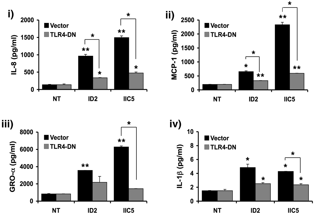
Results: We report that anti-beta(2)GPI antibodies trigger an inflammatory response in trophoblast, characterized by increased secretion of interleukin (IL)-8, MCP-1, GRO-alpha, and IL-1beta, and that this occurs in a TLR-4/MyD88-dependent manner. At high concentrations, these antibodies also induce caspase-mediated cell death. This was attenuated upon disabling of the MyD88 pathway, suggesting that anti-beta(2)GPI-induced inflammatory mediators compromise trophoblast survival by acting in an autocrine/paracrine manner. Enhanced IL-8, GRO-alpha, and IL-1beta secretion also occurred when trophoblast cells were incubated with antibodies from patients with antiphospholipid syndrome. Heparin, which acts as a pro-survival factor in human trophoblast, attenuated the anti-beta(2)GPI antibody-mediated cell death, and also the pro-inflammatory response, but only at high concentrations.

Conclusion: These findings demonstrate that aPL triggers a placental inflammatory response via the TLR-4/MyD88 pathway, which in turn compromises trophoblast survival. Thus, the TLR-4/MyD88 pathway may provide a new therapeutic target to improve pregnancy outcome in antiphospholipid syndrome patients.


Figures

Figure 1
Figure 1. Expression of β2GPI by first trimester trophoblast cells
A) Lysates of the first trimester trophoblast cells (HTR8) and, as a positive control, placental tissue, were analyzed for β2GPI expression by Western blot. The trophoblast cells were positive β2GPI. B) First trimester trophoblast cells (HTR8) were assessed for ID2 and IIC5 binding by immunocytochemistry. Cell were incubated with ID2 or IIC5 (40µg/ml) followed by a peroxidase-conjugated horse anti-mouse antibody (magnification ×60). Note the strong positive immunoreactivity of the trophoblast cells for ID2 and IIC5, localized to both the cytoplasm and plasma membrane. Control cells (Neg; magnification ×40) showed no staining.
Figure 2
Figure 2. Effects of anti-β2GPI Abs on trophoblast cell viability and apoptosis
(A) First trimester trophoblast cells (HTR8) were incubated with ID2, IIC5 or the mouse IgG1 isotype control (IgG) at 0, 5, 10, 20 and 40µg/ml for 48 hours. Cell viability was then determined using the CellTiter 96 assay. Line graph shows the percentage cell viability relative to the untreated control (0 µg/ml). Treatment with ID2 or IIC5 at the high dose of 40µg/ml significantly reduced trophoblast cell viability when compared to the untreated control (*p<0.05, **p<0.001). This figure is representative of at least three independent experiments performed in triplicate. Significance was determined using ANOVA. (B) First trimester trophoblast cells (HTR8) were incubated with no treatment (NT); ID2 (40µg/ml); IIC5 (40µg/ml); mouse IgG (40µg/ml), as a negative isotype control (IgG); or CPT, (4µM) as a positive control for cell death, for 48 hours. Cell viability was then determined using the CellTiter 96 assay. Barchart shows the percentage cell viability relative to the NT control *p <0.05, **p <0.001. Significance was determined using ANOVA. Data are pooled from five individual experiments. (C) First trimester trophoblast cells (HTR8) were incubated with NT, ID2 (40µg/ml); IIC5 (40µg/ml) or mIgG (IgG; 40µg/ml) in the presence and absence of heparin (100ng/ml) for 48 hours. Cell viability was then determined using the CellTiter 96 assay. Barchart shows the percentage cell viability relative to the NT control unless otherwise indicated and significance was determined using ANOVA. The presence of heparin significantly reduced the amount of cell death induced by ID2 and IIC5 (*p <0.01, **p <0.001), as determined using a paired t-test. Data are pooled from three individual experiments. (D) Trophoblast cells (HTR8) were incubated with either no treatment (NT), ID2 (40µg/ml) or IIC5 (40µg/ml). After 48 hours, caspase-3; caspase-8; and caspase-9 activation was determined. Barchart shows caspase activity in relative light units (RLU) relative to the NT control (*p<0.001). Significance was determined using ANOVA. Data are pooled from three individual experiments.
Figure 3
Figure 3. Effects of anti-β2GPI Abs on trophoblast IL-8 secretion
Trophoblast cells (HTR8) were treated with ID2 and IIC5 (0 – 20µg/ml) or the mouse IgG isotype control (mIgG) (20µg/ml) for 72 hours after which cell-free supernatants were collected and analyzed for IL-8 by ELISA. (A) Line graph shows that both ID2 and IIC5 treatment upregulated trophoblast production of IL-8 in a dose dependent manner (*p<0.05, **p<0.001). (B) Barchart shows that while ID2 and IIC5 at 20µg/ml significantly increased trophoblast IL-8 secretion relative the no treatment (NT) control (*p <0.05, **p<0.001), the mIgG had no effect. Significance was determined using ANOVA. (C) Trophoblast cells (HTR8) were incubated with NT, ID2 (20µg/ml) or IIC5 (20µg/ml) in the presence and absence of heparin (10µg/ml) for 72 hours after which cell-free supernatants were collected and analyzed for IL-8 by ELISA. Barchart shows that while ID2 and IIC5 significantly increased trophoblast IL-8 secretion relative the no treatment (NT) control (*p <0.05; **p<0.001), as determined using ANOVA, the presence of heparin significantly inhibited this response as determine by paired t-test (*p <0.05; **p<0.001). Data are representative of at least three independent experiments.
Figure 4
Figure 4. Effects of anti-β2GPI Abs on trophoblast cytokine and chemokine production
(A) HTR8 cells and (B) primary trophoblast cells were treated with either no treatment (NT), ID2 (20µg/ml), IIC5 (20µg/ml) or mouse IgG1 (mIgG; 20µg/ml) for 48 hours after which cell-free supernatants were collected and analyzed by multiplex analysis. Barcharts show changes in the secretion of i) IL-8; ii) MCP-1; iii) GROα; and iv) IL-1β relative to the NT control (*p<0.05; **p<0.001). Data are representative of at least three independent experiments. Significance was determined using ANOVA.
Figure 5
Figure 5. Effects of a TLR4-DN on the modulation of trophoblast cytokine/chemokine production by anti-β2GPI Abs
Trophoblast cells (HTR8) were transiently transfected to express the TLR4-DN. Following transfection with either the TLR4-DN or the vector control, trophoblast cells were treated with either no treatment (NT), ID2 (20µg/ml), or IIC5 (20µg/ml) for 48 hours after which cell-free supernatants were collected and analyzed by multiplex analysis. Barcharts show changes in the secretion of i) IL-8; ii) MCP-1; iii) GROα; and iv) IL-1β. *p<0.05; **p<0.001 relative to the NT control was determined using ANOVA. Significant differences between the TLR4-DN and vector control were determined by paired t-test (*p<0.05; **p<0.001). Data are a representative of at least three independent experiments.
Figure 6
Figure 6. Effects of a MyD88-DN on the modulation of trophoblast cytokine/chemokine production by anti-β2GPI Abs
Trophoblast cells (HTR8) were transiently transfected to express the MyD88-DN. Following transfection with either the MyD88-DN or the vector control, trophoblast cells were treated with either no treatment (NT), ID2 (20µg/ml), or IIC5 (20µg/ml) for 48 hours after which cell-free supernatants were collected and analyzed by multiplex analysis. Barcharts show changes in the secretion of i) IL-8; ii) MCP-1; iii) GROα; and iv) IL-1β. *p<0.05 and **p<0.001 relative to the NT was determined using ANOVA. Significant differences between the MyD88-DN and vector control were determined by paired t-test (*p<0.05; **p<0.001). Data are a representative of at least three independent experiments.
Figure 7
Figure 7. Effects of anti-β2GPI Abs induced inflammation on trophoblast cell death
First trimester trophoblast cells (HTR8) were incubated with no treatment (NT) or the CM from trophoblast cells treated with NT (CM/NT), ID2 at 20µg/ml (CM/ID2) or IIC5 at 20µg/ml (CM/IIC5). After 96 hours, cell viability was determined using the CellTiter 96 assay and after 72 hours caspase-3 activity was determined using the caspase-Glo assay. Barcharts shows (A) the differences in trophoblast cell death; and (B) the differences in trophoblast caspase-3 activity, relative to the NT, as determined using ANOVA (*p <0.05, **p <0.001). Statistical differences between the treatment groups was determined by a paired t-test (*p <0.05, **p <0.001). Data are pooled from three independent experiments. (C) Trophoblast cells (HTR8) were transiently transfected to express the MyD88-DN. Following transfection, trophoblast cells were treated with either no treatment (NT), ID2 (40µg/ml), or IIC5 (40µg/ml) for 48 hours, after which cell viability was determined using the CellTiter 96 assay. Barcharts show the differences in trophoblast cell death relative to the NT control. *p<0.05 and **p<0.001 relative to the NT control was determined using ANOVA. Statistical differences between the MyD88-DN and vector control were determined by a paired t-test (*p <0.05, **p <0.001). Data are pooled from three independent experiments.
Figure 8
Figure 8. Effects of patient derived aPL on trophoblast cytokine/chemokine production
Trophoblast cells were incubated with either no treatment (NT) or polyclonal IgG aPL (12.5µg/ml) from patients with PM+/VT (n=6); PM/VT+ (n=6) or PM+/VT+ (n=6). After 72 hours, the supernatants were collected, pooled on the basis of patient group, and than evaluated by multiplex analysis. Barcharts show the effects of aPL on trophoblast secretion of i) IL-8; ii) MCP-1; iii) GROα; and iv) IL-1β after 72 hours. *p<0.05 relative to the NT control, as determined using ANOVA.

Similar articles

Cited by

References

    1. D'Cruz DP, Khamashta MA, Hughes GR. Systemic lupus erythematosus. Lancet. 2007;369:587–596. - PubMed
    1. Valesini G, Alessandri C. New facet of antiphospholipid antibodies. Ann N Y Acad Sci. 2005;1051:487–497. - PubMed
    1. Rai RS, Regan L, Clifford K, Pickering W, Dave M, Mackie I, McNally T, Cohen H. Antiphospholipid antibodies and beta 2-glycoprotein-I in 500 women with recurrent miscarriage: results of a comprehensive screening approach. Hum Reprod. 1995;10:2001–2005. - PubMed
    1. Regan L, Rai R. Epidemiology and the medical causes of miscarriage. Baillieres Best Pract Res Clin Obstet Gynaecol. 2000;14:839–854. - PubMed
    1. Cervera R, Piette JC, Font J, Khamashta MA, Shoenfeld Y, Camps MT, Jacobsen S, Lakos G, Tincani A, Kontopoulou-Griva I, Galeazzi M, Meroni PL, Derksen RH, de Groot PG, Gromnica-Ihle E, Baleva M, Mosca M, Bombardieri S, Houssiau F, Gris JC, Quere I, Hachulla E, Vasconcelos C, Roch B, Fernandez-Nebro A, Boffa MC, Hughes GR, Ingelmo M. Antiphospholipid syndrome: clinical and immunologic manifestations and patterns of disease expression in a cohort of 1,000 patients. Arthritis Rheum. 2002;46:1019–1027. - PubMed

Publication types

MeSH terms