Neonatal exposure to parathion alters lipid metabolism in adulthood: Interactions with dietary fat intake and implications for neurodevelopmental deficits
- PMID: 19615431
- PMCID: PMC2795103
- DOI: 10.1016/j.brainresbull.2009.07.002
Neonatal exposure to parathion alters lipid metabolism in adulthood: Interactions with dietary fat intake and implications for neurodevelopmental deficits
Abstract
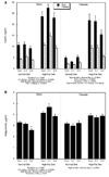
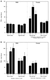
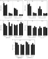
Organophosphates are developmental neurotoxicants but recent evidence also points to metabolic dysfunction. We determined whether neonatal parathion exposure in rats has long-term effects on regulation of adipokines and lipid peroxidation. We also assessed the interaction of these effects with increased fat intake. Rats were given parathion on postnatal days 1-4 using doses (0.1 or 0.2mg/kg/day) that straddle the threshold for barely detectable cholinesterase inhibition and the first signs of systemic toxicity. In adulthood, animals were either maintained on standard chow or switched to a high-fat diet for 7 weeks. We assessed serum leptin and adiponectin, tumor necrosis factor-alpha (TNFalpha) in adipose tissues, and thiobarbituric acid reactive species (TBARS) in peripheral tissues and brain regions. Neonatal parathion exposure uncoupled serum leptin levels from their dependence on body weight, suppressed adiponectin and elevated TNFalpha in white adipose tissue. Some of the effects were offset by a high-fat diet. Parathion reduced TBARS in the adipose tissues, skeletal muscle and temporal/occipital cortex but not in heart, liver, kidney or frontal/parietal cortex; it elevated TBARS in the cerebellum; the high-fat diet again reversed many of the effects. Neonatal parathion exposure disrupts the regulation of adipokines that communicate metabolic status between adipose tissues and the brain, while also evoking an inflammatory adipose response. Our results are consistent with impaired fat utilization and prediabetes, as well as exposing a potential relationship between effects on fat metabolism and on synaptic function in the brain.
Conflict of interest statement
Conflict of Interest
The authors do not have any conflicts of interest. However, TAS has provided expert witness testimony in the past three years at the behest of the following law firms: The Calwell Practice (Charleston WV), Frost Brown Todd (Charleston WV), Snyder Weltchek & Snyder (Baltimore MD), Finnegan Henderson Farabow Garrett & Dunner (Washington DC), Frommer Lawrence Haug (Washington DC), Carter Law (Peoria IL), Corneille Law (Madison WI), Angelos Law (Baltimore MD), Kopff, Nardelli & Dopf (New York NY), and Gutglass Erickson Bonville & Larson (Madison WI).
Figures


Similar articles
-
Neonatal parathion exposure and interactions with a high-fat diet in adulthood: Adenylyl cyclase-mediated cell signaling in heart, liver and cerebellum.Brain Res Bull. 2010 Apr 5;81(6):605-12. doi: 10.1016/j.brainresbull.2010.01.003. Epub 2010 Jan 14. Brain Res Bull. 2010. PMID: 20074626 Free PMC article.
-
Exposure of neonatal rats to parathion elicits sex-selective reprogramming of metabolism and alters the response to a high-fat diet in adulthood.Environ Health Perspect. 2008 Nov;116(11):1456-62. doi: 10.1289/ehp.11673. Epub 2008 Jun 23. Environ Health Perspect. 2008. PMID: 19057696 Free PMC article.
-
Consumption of a high-fat diet in adulthood ameliorates the effects of neonatal parathion exposure on acetylcholine systems in rat brain regions.Environ Health Perspect. 2009 Jun;117(6):916-22. doi: 10.1289/ehp.0800459. Epub 2009 Feb 3. Environ Health Perspect. 2009. PMID: 19590683 Free PMC article.
-
Neonatal parathion exposure disrupts serotonin and dopamine synaptic function in rat brain regions: modulation by a high-fat diet in adulthood.Neurotoxicol Teratol. 2009 Nov-Dec;31(6):390-9. doi: 10.1016/j.ntt.2009.07.003. Epub 2009 Jul 16. Neurotoxicol Teratol. 2009. PMID: 19616088 Free PMC article.
-
Does early-life exposure to organophosphate insecticides lead to prediabetes and obesity?Reprod Toxicol. 2011 Apr;31(3):297-301. doi: 10.1016/j.reprotox.2010.07.012. Epub 2010 Sep 17. Reprod Toxicol. 2011. PMID: 20850519 Free PMC article. Review.
Cited by
-
The Effects of Endocrine Disruptors on Adipogenesis and Osteogenesis in Mesenchymal Stem Cells: A Review.Front Endocrinol (Lausanne). 2017 Jan 9;7:171. doi: 10.3389/fendo.2016.00171. eCollection 2016. Front Endocrinol (Lausanne). 2017. PMID: 28119665 Free PMC article. Review.
-
Neonatal parathion exposure and interactions with a high-fat diet in adulthood: Adenylyl cyclase-mediated cell signaling in heart, liver and cerebellum.Brain Res Bull. 2010 Apr 5;81(6):605-12. doi: 10.1016/j.brainresbull.2010.01.003. Epub 2010 Jan 14. Brain Res Bull. 2010. PMID: 20074626 Free PMC article.
-
Pesticide use and incident diabetes among wives of farmers in the Agricultural Health Study.Occup Environ Med. 2014 Sep;71(9):629-35. doi: 10.1136/oemed-2013-101659. Epub 2014 Apr 12. Occup Environ Med. 2014. PMID: 24727735 Free PMC article.
-
High fat diet intake during pre and periadolescence impairs learning of a conditioned place preference in adulthood.Behav Brain Funct. 2011 Jun 26;7:21. doi: 10.1186/1744-9081-7-21. Behav Brain Funct. 2011. PMID: 21703027 Free PMC article.
-
Paraoxonase 1 polymorphism and prenatal pesticide exposure associated with adverse cardiovascular risk profiles at school age.PLoS One. 2012;7(5):e36830. doi: 10.1371/journal.pone.0036830. Epub 2012 May 15. PLoS One. 2012. PMID: 22615820 Free PMC article.
References
-
- Abdollahi M, Donyavi M, Pournourmohammadi S, Saadat M. Hyperglycemia associated with increased hepatic glycogen phosphorylase and phosphoenolpyruvate carboxykinase in rats following subchronic exposure to malathion. Comp. Biochem. Physiol. Toxicol. Pharmacol. 2004;137:343–347. - PubMed
-
- Ahima RS. Adipose tissue as an endocrine organ. Obesity. 2006;14:242S–249S. - PubMed
-
- Alexandraki K, Piperi C, Kalofoutis C, Singh J, Alaveras A, Kalofoutis A. Inflammatory process in type 2 diabetes: the role of cytokines. Ann. N.Y. Acad. Sci. 2006;1084:89–117. - PubMed
-
- Beltowski J, Wojcicka G, Gorny D, Marciniak A. The effect of dietary-induced obesity on lipid peroxidation, antioxidant enzymes and total plasma antioxidant capacity. J. Physiol. Pharmacol. 2000;51:883–896. - PubMed
-
- Casida JE, Quistad GB. Organophosphate toxicology: safety aspects of nonacetylcholinesterase secondary targets. Chem. Res. Toxicol. 2004;17:983–998. - PubMed
Publication types
MeSH terms
Substances
Grants and funding
LinkOut - more resources
Full Text Sources
Medical

