. 2009 Sep 25;387(3):521-4.
doi: 10.1016/j.bbrc.2009.07.060. Epub 2009 Jul 16.

Carbachol induces p70S6K1 activation through an ERK-dependent but Akt-independent pathway in human colonic epithelial cells

Affiliations

Carbachol induces p70S6K1 activation through an ERK-dependent but Akt-independent pathway in human colonic epithelial cells

Xiaohua Jiang et al. Biochem Biophys Res Commun. .

Abstract

Stimulation of human colonic epithelial T84 cells with the muscarinic receptor agonist carbachol, a stable analog of acetylcholine, induced Akt, p70S6K1 and ERK activation. Treatment of T84 cells with the selective inhibitor of EGF receptor (EGFR) tyrosine kinase AG1478 abrogated Akt phosphorylation on Ser(473) induced by either carbachol or EGF, indicating that carbachol-induced Akt activation is mediated through EGFR transactivation. Surprisingly, AG1478 did not suppress p70S6K1 phosphorylation on Thr(389) in response to carbachol, indicating the G protein-coupled receptor (GPCR) stimulation induces p70S6K1 activation, at least in part, via an Akt-independent pathway. In contrast, treatment with the selective MEK inhibitor U0126 (but not with the inactive analog U0124) inhibited carbachol-induced p70S6K1 activation, indicating that the MEK/ERK/RSK pathway plays a critical role in p70S6K1 activation in GPCR-stimulated T84 cells. These findings imply that GPCR activation induces p70S6K1 via ERK rather than through the canonical PI 3-kinase/Akt/TSC/mTORC1 pathway in T84 colon carcinoma cells.


Figures
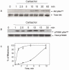
Figure 1
Figure 1. Carbachol induces Akt phosphorylation on Ser473 and p70S6K phosphorylation on Ser389in a time-dependent fashion in T84 cells
Cultures of T84 cells grown on 35 mm dishes were stimulated with 100 μM carbachol for the indicated times. All cultures were lysed with 2×SDS-PAGE sample buffer and analyzed by SDS-PAGE and immunoblotting with antibodies that detect either Akt phosphorylated on Ser473 (panel A) or p70S6K phosphorylated on Ser389 (panel B). Antibodies that detect total Akt or p70S6K were used to verify equivalent loading of the gel. Autoluminograms were quantified by densitometric scanning. The results shown are the mean ± S.E.M. n=3 and are expressed as percentage of the maximum increase induced by treatment with carbachol for either AKT pSer473 closed symbols or p70S6K pSer389 open symbols (panel C).
Figure 2
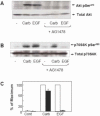
Figure 2. Carbachol induced Akt phosphorylation on Ser473 is EGFR-dependent but p70S6K phosphorylation on Ser389 is EGFR-independent
A and B. T84 cells were incubated in the absence or presence of the EGFR inhibitor AG1478 (1 μM) for 1 h prior to stimulation of the cells with 100 μM carbachol (carb) or 50 ng/ml EGF for 10 min. The cultures were lysed with 2×SDS-PAGE sample buffer and analyzed by SDS-PAGE and immunoblotting with phospho Akt Ser473 and total Akt in panel A and phospho p70S6K Ser389 and total p70S6K to verify equal loading in panel B. Shown here are representative autoluminograms; similar results were obtained in 3 independent experiments. Autoluminograms were quantified by densitometric scanning. The results shown are the mean ± S.E.M. n=3 and are expressed as percentage of the maximum increase induced by treatment with carbachol, in cells preincubated in the absence (open bars) or the presence (closed bars) of AG1478 (panel C).
Figure 3
Figure 3. Carbachol induced p70S6K phosphorylation through an ERK-dependent pathway
A. Cultures of T84 cells were stimulated with 100 μM carbachol for the indicated times, lysed with 2×SDS-PAGE sample buffer and analyzed by SDS-PAGE and immunoblotting with phospho-ERK antibody (pERK1/2). Shown here are representative autoluminograms; similar results were obtained in 3 independent experiments. B, T84 cells were incubated in the absence or presence of the MEK1/2 inhibitor U0126 (10 μM) or the inactive analogue U0124 for 1 h prior to stimulation of the cells with 100 μM carbachol (carb) for 10 min. The cultures were lysed with 2×SDS-PAGE sample buffer and analyzed by SDS-PAGE and immunoblotting with the following antibodies: phospho ERK1/2, p70S6K Ser389 and total p70S6K. Shown here are representative autoluminograms; similar results were obtained in 3 independent experiments.

Similar articles

Cited by

References

    1. Rozengurt E. Early signals in the mitogenic response. Science. 1986;234:161–166. - PubMed
    1. Rozengurt E. Neuropeptides as growth factors for normal and cancer cells. Trends Endocrinol Metabol. 2002;13:128–134. - PubMed
    1. Rozengurt E, Walsh JH. Gastrin, CCK, signaling, and cancer. Annu. Rev. Physiol. 2001;63:49–76. - PubMed
    1. Rozengurt E. Mitogenic signaling pathways induced by G protein-coupled receptors. J Cell Physiol. 2007;213:589–602. - PubMed
    1. Rozengurt E. Growth factors and cell proliferation. Curr. Opin. Cell Biol. 1992;4:161–165. - PubMed

Publication types

MeSH terms