Review
. 2009 Oct;47(4):436-44.
doi: 10.1016/j.yjmcc.2009.07.008. Epub 2009 Jul 16.

Molecular and biophysical mechanisms of Ca2+ sparklets in smooth muscle

Affiliations
Review

Molecular and biophysical mechanisms of Ca2+ sparklets in smooth muscle

Luis F Santana et al. J Mol Cell Cardiol. 2009 Oct.

Abstract

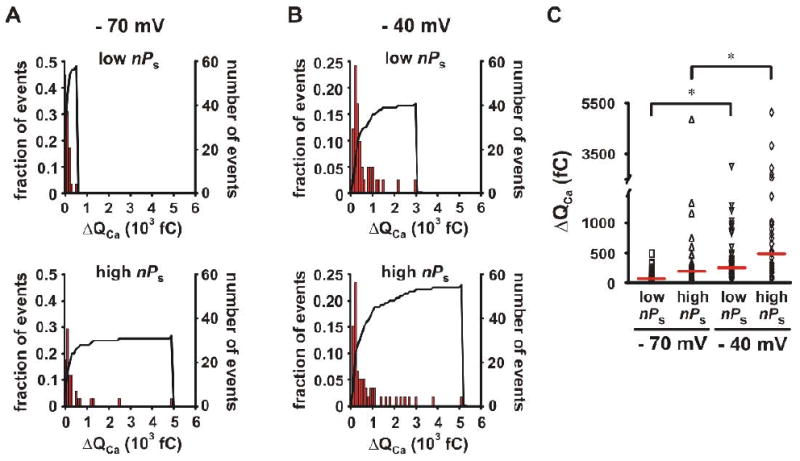
In this article, we review the biophysical basis and functional implications of a novel Ca(2+) signal (called "Ca(2+) sparklets") produced by Ca(2+) influx via L-type Ca(2+) channels (LTCCs) in smooth muscle. Ca(2+) sparklet activity is bimodal. In low activity mode, Ca(2+) sparklets are produced by random, brief openings of solitary LTCCs. In contrast, small clusters of LTCCs can function in a high activity mode that creates sites of continual Ca(2+) influx called "persistent Ca(2+) sparklets". Low activity and persistent Ca(2+) sparklets contribute to Ca(2+) influx in arterial, colonic, and venous smooth muscle. Targeting of PKCalpha by the scaffolding protein AKAP150 to specific sarcolemmal domains is required for the activation of persistent Ca(2+) sparklets. Calcineurin, which is also associated with AKAP150, opposes the actions of PKCalpha on Ca(2+) sparklets. At hyperpolarized potentials, Ca(2+) sparklet activity is low and hence does not contribute to global [Ca(2+)](i). Membrane depolarization increases low and persistent Ca(2+) sparklet activity, thereby increasing local and global [Ca(2+)](i). Ca(2+) sparklet activity is increased in arterial myocytes during hypertension, thus increasing Ca(2+) influx and activating the transcription factor NFATc3. We discuss a model for subcellular variations in Ca(2+) sparklet activity and their role in the regulation of excitation-contraction coupling and excitation-transcription coupling in smooth muscle.


Figures

Figure 1
Figure 1. Ca2+ sparklets in arterial and venous myocytes
(A) TIRF images of Ca2+ sparklets from representative arterial (left) and portal vein (right) smooth muscle cells (-70 mV; external Ca2+ = 20 mM). Traces below each image show the time-course of [Ca2+]i in the sites marked by the green circles. (B) Simultaneous recordings of Ca2+ sparklets and unitary LTCC currents. Redrawn from [8].
Figure 2
Figure 2. Ca2+ sparklet activity is quantal
All-points histogram generated from 25 representatives blank TIRF traces (A) or Ca2+ sparklet traces (B) similar to those in the inserts. (C) Description of Ca2+ sparklet nPs analysis. Solid lines represent fit of the [Ca2+]i traces using the single channel detection and analysis algorithms of Clampex 9.0. Dotted lines represent quantal levels. Figure redrawn from [9].
Figure 3
Figure 3. Proposed model for heterogeneous Ca2+ sparklet activity and NFATc3 activation in arterial myocytes
Activation of hetero-trimeric Gq/11 proteins by a membrane receptor like the angiotensin receptor type 1 (AT1R) activates phospholipase C (PLC), which increases diacylglycerol (DAG) levels and activates PKCα AKAP150 targets PKCα and calcineurin to specific regions of the sarcolemma where they can modulate the activity of nearby LTCCs. Ca2+ entering the cell recruits calmodulin, thus releasing PKCα from its interaction with AKAP150 allowing it to phosphorylate neighboring LTCCs. LTCCs associated with the AKAP150/PKCα/calcineurin complex can develop persistent Ca2+ sparklet activity, forming a signaling unit that modulates Ca2+ influx and NFATc3-dependent gene expression in smooth muscle. Initiation or continuation of this signaling unit could be prevented by inhibiting PKCα, calcineurin, or NFATc3 activation or with L-type Ca2+ channel blockers. Green arrows indicate activation; red arrows indicate inhibition/down-regulation.
Figure 4
Figure 4. Signal mass of Ca2+ sparklets in smooth muscle
Amplitude histograms of ΔQCa from low and high nPs Ca2+ sparklet sites at −70 (A) and −40 mV (B). (C) Scatter plot of ΔQCa amplitudes from low and high nPs Ca2+ sparklet sites at −70 and −40 mV; the red line represents the median value for each column of values. Figure redrawn from [10].
Figure 5
Figure 5. The effects of blocking Ca2+ sparklets on global [Ca2+]i at -70 mV
Simulation of the effects of blocking persistent Ca2+ sparklets on global [Ca2+]i, measured in a cell loaded with Fluo-5F using epifluorescence (A) or TIRF (B) microscopy. Right side panels show all-points histograms from the [Ca2+]i records undergoing a decrease of 14 nM [Ca2+]i. Solid lines in the histograms represent the best fit of the data using a Gaussian function.

Similar articles

Cited by

References

    1. Nieves-Cintrón M, Amberg GC, Navedo MF, Molkentin JD, Santana LF. The control of Ca2+ influx and NFATc3 signaling in arterial smooth muscle during hypertension. Proc Natl Acad Sci U S A. 2008;105(40):15623–8. - PMC - PubMed
    1. Amberg GC, Rossow CF, Navedo MF, Santana LF. NFATc3 regulates Kv2.1 expression in arterial smooth muscle. J Biol Chem. 2004;279(45):47326–34. - PubMed
    1. Cartin L, Lounsbury KM, Nelson MT. Coupling of Ca(2+) to CREB activation and gene expression in intact cerebral arteries from mouse : roles of ryanodine receptors and voltage-dependent Ca(2+) channels. Circ Res. 2000;86(7):760–7. - PubMed
    1. Knot HJ, Nelson MT. Regulation of arterial diameter and wall [Ca2+] in cerebral arteries of rat by membrane potential and intravascular pressure. J Physiol. 1998;508(Pt 1):199–209. - PMC - PubMed
    1. Harder DR, Gilbert R, Lombard JH. Vascular muscle cell depolarization and activation in renal arteries on elevation of transmural pressure. Am J Physiol. 1987 Oct;253(4 Pt 2):F778–81. - PubMed

Publication types