Review
. 2009 Aug;30(8):431-40.
doi: 10.1016/j.tips.2009.05.005. Epub 2009 Jul 16.

PET radiotracers: crossing the blood-brain barrier and surviving metabolism

Affiliations
Review

PET radiotracers: crossing the blood-brain barrier and surviving metabolism

Victor W Pike. Trends Pharmacol Sci. 2009 Aug.

Abstract

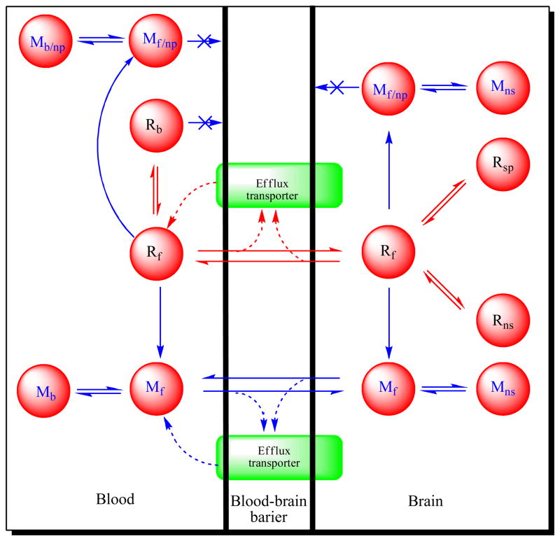
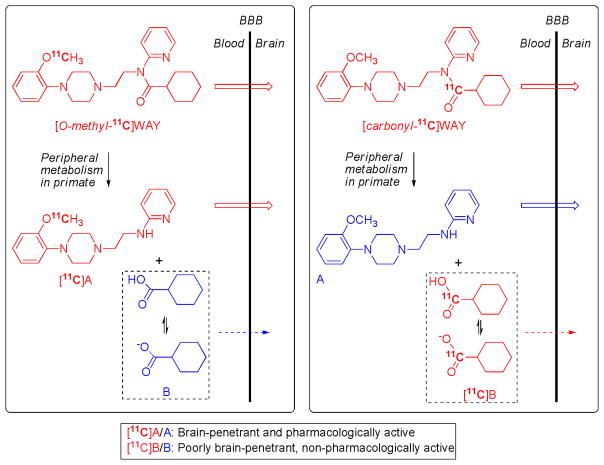
Radiotracers for imaging protein targets in the living human brain with positron emission tomography (PET) are increasingly useful in clinical research and in drug development. Such radiotracers must fulfill many criteria, among which an ability to enter brain adequately and reversibly without contamination by troublesome radiometabolites is desirable for accurate measurement of the density of a target protein (e.g. neuroreceptor, transporter, enzyme or plaque). Candidate radiotracers can fail as a result of poor passive brain entry, rejection from brain by efflux transporters or undesirable metabolism. These issues are reviewed. Emerging PET radiotracers for measuring efflux transporter function and new strategies for ameliorating radiotracer metabolism are discussed. A growing understanding of the molecular features affecting the brain penetration, metabolism and efflux transporter sensitivity of prospective radiotracers should ultimately lead to their more rational and efficient design, and also to their greater efficacy.


Figures

Figure 1
Figure 1
PET Radiotracers: crossing the blood-brain barrier and surviving metabolism. The cartoon represents a voxel in brain, with edge dimensions of a millimeter or so, as seen with a PET camera after intravenous administration of a radiotracer. Blood represents about 5% of the total volume. The red circles indicate some of the radioactive entities that might be generated from the parent radiotracer (Rf) and their locations. The green areas represent efflux transporters at the blood-brain barrier (e.g., P-gp). Key: R = radiotracer; M = radiometabolite. Subscripts: f = free; b = plasma protein-bound; np = non BBB-penetrant; ns = non-specifically bound to brain; sp = specifically bound to brain. Radiotracer development for a protein target within brain aims to achieve ready passive diffusion of the radiotracer across the BBB, without impediment by efflux transporters, to give a high ratio of specifically bound radiotracer (Rsp) to non-specifically bound (Rnp) plus free (Rf) radiotracer, while suppressing the presence of radiometabolite entities in brain.
Figure 2
Figure 2
[11C]WAY-110635 for imaging brain 5-HT1A receptors: avoiding troublesome radiometabolites by judicious choice of position of radiolabel. WAY-100635 is metabolized in the periphery by hydrolysis of its amide bond. When WAY-100635 is labeled with carbon-11 in its O-methyl group, metabolism produces [11C]A as a major radiometabolite in plasma. [11C]A is able to enter brain to bind non-specifically, and also potentially specifically to some proteins for which it has moderately high affinity (5-HT1A receptors and α1 adrenoceptors) [54]. This radiometabolite therefore attenuates the signal from the unchanged radiotracer, [O-methyl-11C]WAY-100635, and exacerbates biomathematical analysis. By placing the radiolabel in the carbonyl position, metabolism of the radiotracer, [carbonyl-11C]WAY-100635, to [11C]A is avoided. The major radiometabolite becomes [11C]cyclohexanecarboxylate anion, which has only low transient entry into brain [55]. Consequently, with this radiotracer a much greater proportion of the radioactivity in brain is specifically bound unchanged radioligand [27], and biomathematical modeling of the acquired PET data is possible to deliver measures of receptor density [79, 80].

References

    1. Jacobs AH, et al. PET-based molecular imaging in neuroscience. Eur J Nucl Med & Mol Imaging. 2003;30:1051–1065. - PubMed
    1. Lee CM, Farde L. Using positron emission tomography to facilitate drug development. TiPs. 2006;27:310–316. - PubMed
    1. Fowler JS, Ido T. In: Handbook of Radiopharmaceuticals, Radiochemistry and Applications. Welch MJ, Redvanly CS, editors. Chapter 9. Wiley; Chichester: 2003. pp. 307–321.
    1. Pike VW. Positron-emitting radioligands for studies in vivo Probes for human psychopharmacology. J Psychopharmacology. 1993;7:139–158. - PubMed
    1. Laruelle M, et al. Relationships between radiotracer properties and image quality in molecular imaging of the brain with positron emission tomography. Mol Imaging Biol. 2003;5:363–375. - PubMed

Publication types

MeSH terms