. 2009 Sep 18;392(2):452-64.
doi: 10.1016/j.jmb.2009.07.028. Epub 2009 Jul 17.

Highly cooperative recruitment of Ets-1 and release of autoinhibition by Pax5

Affiliations

Highly cooperative recruitment of Ets-1 and release of autoinhibition by Pax5

Daniel Fitzsimmons et al. J Mol Biol. .

Abstract

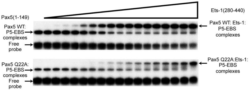
Pax5 (paired box binding factor 5) is a critical regulator of transcription and lineage commitment in B lymphocytes. In B cells, mb-1 (Ig-alpha/immunoglobulin-associated alpha) promoter transcription is activated by Pax5 through its recruitment of E74-like transforming sequence (Ets) family proteins to a composite site, the P5-EBS (Pax5-Ets binding site). Previously, X-ray crystallographic analysis revealed a network of contacts between the DNA-binding domains of Pax5 and Ets-1 while bound to the P5-EBS. Here, we report that Pax5 assembles these ternary complexes via highly cooperative interactions that overcome the autoinhibition of Ets-1. Using recombinant proteins, we calculated K(d(app)) values for the binding of Pax5, Ets-1, and GA-binding proteins, separately or together, to the P5-EBS. By itself, Pax5 binds the P5-EBS with high affinity (K(d) approximately equal 2 nM). Ets-1(331-440) bound the P5-EBS by itself with low affinity (K(d)=136 nM). However, autoinhibited Ets-1(280-440) alone does not bind detectably to the suboptimal sequences of the P5-EBS. Recruitment of Ets-1(331-440) or Ets-1(280-440) resulted in highly efficient ternary complex assembly with Pax5. Pax5 counteracts autoinhibition and increases binding of Ets-1 of the mb-1 promoter by >1000-fold. Mutation of Pax5 Gln22 to alanine (Q22A) enhances promoter binding by Pax5; however, Q22A greatly reduces recruitment of Ets-1(331-440) and Ets-1(280-440) by Pax5 (8.9- or >300-fold, respectively). Thus, Gln22 of Pax5 is essential for overcoming Ets-1 autoinhibition. Pax5 wild type and Q22A each recruited GA-binding protein alpha/beta1 to the mb-1 promoter with similar affinities, but recruitment was less efficient than that of Ets-1 (reduced by approximately 8-fold). Our results suggest a mechanism that allows Pax5 to overcome autoinhibition of Ets-1 DNA binding. In summary, these data illustrate requirements for partnerships between Ets proteins and Pax5.


Figures

Fig. 1
Fig. 1
Recombinant proteins used in these studies. Proteins utilized in these studies include human Pax5(1–149) and murine Ets-1(280–440), Ets-1(331–440), GABPα (311–420) and GABPβ1(1–157). Open boxes representα–helices. Arrows represent β-sheets. Filled boxes denote the Notch-like/ankyrin repeats of GABPβ1. Boundaries of the paired domain of Pax5 (15–143) and ETS domains of Ets-1 and GABPα are indicated. The autoinhibitory domains of Ets-1 (shaded) and residues that are highlighted in the text are also indicated.
Fig. 2
Fig. 2
Pax5(1–149)WT or Q22A bind similarly to the P5-EBS probe. (a) Representative equilibrium DNA binding analysis by EMSA with increasing amounts of Pax5 (1–149) WT (upper panel) or Q22A (lower panel) and a constant amount of the P5-EBS probe. (b) Non-linear least squares analysis of DNA binding complexes in (a). Protein concentrations shown in the graph were corrected for relative percent functional activities.
Fig. 2
Fig. 2
Pax5(1–149)WT or Q22A bind similarly to the P5-EBS probe. (a) Representative equilibrium DNA binding analysis by EMSA with increasing amounts of Pax5 (1–149) WT (upper panel) or Q22A (lower panel) and a constant amount of the P5-EBS probe. (b) Non-linear least squares analysis of DNA binding complexes in (a). Protein concentrations shown in the graph were corrected for relative percent functional activities.
Fig. 3
Fig. 3
Ets-1(331–440), but not Ets-1(280–440), binds to the P5-EBS probe. (a) Representative equilibrium DNA binding analysis by EMSA with increasing amounts of Ets-1(331–440) (upper panel) or (280–440) (lower panel) and a constant amount of the P5-EBS probe. (b) Non-linear least squares analysis of DNA binding complexes in (a). Graphs were adjusted for active protein concentrations as in Fig. 2b.
Fig. 3
Fig. 3
Ets-1(331–440), but not Ets-1(280–440), binds to the P5-EBS probe. (a) Representative equilibrium DNA binding analysis by EMSA with increasing amounts of Ets-1(331–440) (upper panel) or (280–440) (lower panel) and a constant amount of the P5-EBS probe. (b) Non-linear least squares analysis of DNA binding complexes in (a). Graphs were adjusted for active protein concentrations as in Fig. 2b.
Fig. 4
Fig. 4
Pax5 recruitment of Ets-1(331–440) or Ets-1(280–440) to bind the P5-EBS probe is highly cooperative and requires Gln22 of Pax5. (a) Representative equilibrium DNA binding analysis by EMSA with a constant amount of Pax5(1–149) WT (upper panel) or Q22A (lower panel), increasing amounts of Ets-1(331–440) and a constant amount of the P5-EBS probe. (b) Non-linear least squares analysis of DNA binding complexes in (A). Graphs were adjusted for active protein concentrations as in Fig. 2b. (c) Representative equilibrium DNA binding analysis by EMSA with a constant amount of Pax5(1–149) WT (upper panel) or Q22A (lower panel), increasing amounts of Ets-1(280–440) and a constant amount of the P5-EBS probe. (d) Nonlinear least squares analysis of DNA binding complexes in (c). Graphs were adjusted for active protein concentrations as in Fig. 2b.
Fig. 4
Fig. 4
Pax5 recruitment of Ets-1(331–440) or Ets-1(280–440) to bind the P5-EBS probe is highly cooperative and requires Gln22 of Pax5. (a) Representative equilibrium DNA binding analysis by EMSA with a constant amount of Pax5(1–149) WT (upper panel) or Q22A (lower panel), increasing amounts of Ets-1(331–440) and a constant amount of the P5-EBS probe. (b) Non-linear least squares analysis of DNA binding complexes in (A). Graphs were adjusted for active protein concentrations as in Fig. 2b. (c) Representative equilibrium DNA binding analysis by EMSA with a constant amount of Pax5(1–149) WT (upper panel) or Q22A (lower panel), increasing amounts of Ets-1(280–440) and a constant amount of the P5-EBS probe. (d) Nonlinear least squares analysis of DNA binding complexes in (c). Graphs were adjusted for active protein concentrations as in Fig. 2b.
Fig. 5
Fig. 5
Binding of the P5-EBS by GABPα is dependent on GABPβ1. (a) Representative equilibrium DNA binding analysis by EMSA with increasing amounts of GABPα/β1 (upper panel) or GABPα (lower panel) and a constant amount of the P5-EBS probe. (b) Non-linear least squares analysis of DNA binding complexes in (a). Graphs were adjusted for active protein concentrations as in Fig. 2b.
Fig. 5
Fig. 5
Binding of the P5-EBS by GABPα is dependent on GABPβ1. (a) Representative equilibrium DNA binding analysis by EMSA with increasing amounts of GABPα/β1 (upper panel) or GABPα (lower panel) and a constant amount of the P5-EBS probe. (b) Non-linear least squares analysis of DNA binding complexes in (a). Graphs were adjusted for active protein concentrations as in Fig. 2b.
Fig. 6
Fig. 6
Cooperative binding of the P5-EBS by Pax5 and GABPα or GABPα/β1 does not require Gln22 of Pax5. (a) Representative equilibrium DNA binding analysis by EMSA with a constant amount of Pax5(1–149) WT (upper panel) or Pax5 Q22A (lower panel), increasing amounts of GABPα and a constant amount of the P5-EBS probe. (b) Non-linear least squares analysis of DNA binding complexes in (a). Graphs were adjusted for active protein concentrations as in Fig. 2b. (c) Representative EMSA with a constant amount of Pax5(1–149) WT (upper panel) or Q22A (lower panel), increasing amounts of GABPα/β1 complexes and a constant amount of the P5-EBS probe. (d) Non-linear least squares analysis of DNA binding complexes in (c). Graphs were adjusted for active protein concentrations as in Fig. 2b.
Fig. 6
Fig. 6
Cooperative binding of the P5-EBS by Pax5 and GABPα or GABPα/β1 does not require Gln22 of Pax5. (a) Representative equilibrium DNA binding analysis by EMSA with a constant amount of Pax5(1–149) WT (upper panel) or Pax5 Q22A (lower panel), increasing amounts of GABPα and a constant amount of the P5-EBS probe. (b) Non-linear least squares analysis of DNA binding complexes in (a). Graphs were adjusted for active protein concentrations as in Fig. 2b. (c) Representative EMSA with a constant amount of Pax5(1–149) WT (upper panel) or Q22A (lower panel), increasing amounts of GABPα/β1 complexes and a constant amount of the P5-EBS probe. (d) Non-linear least squares analysis of DNA binding complexes in (c). Graphs were adjusted for active protein concentrations as in Fig. 2b.
Fig. 7
Fig. 7
Model of interaction between the paired domain of Pax5 and Ets-1(280–440) on mb-1 promoter DNA. (a) Overview of the ternary complex. The paired domain of Pax5 is represented in blue, the ETS domain of Ets-1 in magenta and the inhibitory regions of Ets-1 in green. Helix HI-1 of Ets-1 is shown in its unfolded state. (b) Enlarged view of the interface between Pax5 and Ets-1 on mb-1 promoter DNA. Hydrogen bonds between Gln22 of Pax5 and Gln336 or Tyr395 are indicated by dashed lines. A dashed line also indicates proposed interactions between H1 of Ets-1 and the sugar-phosphate backbone of DNA. The figure was made using VMD.
Fig. 7
Fig. 7
Model of interaction between the paired domain of Pax5 and Ets-1(280–440) on mb-1 promoter DNA. (a) Overview of the ternary complex. The paired domain of Pax5 is represented in blue, the ETS domain of Ets-1 in magenta and the inhibitory regions of Ets-1 in green. Helix HI-1 of Ets-1 is shown in its unfolded state. (b) Enlarged view of the interface between Pax5 and Ets-1 on mb-1 promoter DNA. Hydrogen bonds between Gln22 of Pax5 and Gln336 or Tyr395 are indicated by dashed lines. A dashed line also indicates proposed interactions between H1 of Ets-1 and the sugar-phosphate backbone of DNA. The figure was made using VMD.

Similar articles

Cited by

References

    1. Hollenhorst PC, Jones DA, Graves BJ. Expression profiles frame the promoter specificity dilemma of the ETS family of transcription factors. Nucl Acids Res. 2004;32:5693–5702. - PMC - PubMed
    1. Karim FD, Urness LD, Thummel CS, Klemsz MJ, McKercher SR, Celada A, Beveran Cv, Maki RA, Gunther CV, Nye JA, Graves BJ. The ETS-domain: a new DNA-binding motif that recognizes a purine-rich core DNA sequence. Genes Dev. 1990;4:1451–1453. - PubMed
    1. Thompson CC, Brown TA, McKnight SL. Convergence of ets- and notch-related structural motifs in a heteromeric DNA binding complex. Science. 1991;253:762–768. - PubMed
    1. Batchelor AH, Piper DE, de la Brousse FC, McKnight SL, Wolberger C. The structure of GABPα/β: an ETS domain-ankyrin repeat heterodimer bound to DNA. Science. 1998;279:1037–1041. - PubMed
    1. Chinenov Y, Henzl M, Martin ME. The α and β subunits of the GA-binding protein form a stable heterodimer in solution. J Biol Chem. 2000;275:7749–7756. - PubMed

Publication types

MeSH terms