Transmission and expansion of HOXB4-induced leukemia in two immunosuppressed dogs: implications for a new canine leukemia model
- PMID: 19616601
- PMCID: PMC2748853
- DOI: 10.1016/j.exphem.2009.07.004
Transmission and expansion of HOXB4-induced leukemia in two immunosuppressed dogs: implications for a new canine leukemia model
Abstract
Objective: There are currently no large animal models to study the biology of leukemia and development of novel antileukemia therapies. We have previously shown that dogs transplanted with homeobox B4 (HOXB4)-transduced autologous CD34(+) cells developed myeloid leukemia associated with HOXB4 overexpression. Here we describe the transmission, engraftment, and expansion of these canine leukemia cells into two genetically unrelated, immunosuppressed dogs.
Materials and methods: Two dogs immunosuppressed after major histocompatibility complex-haploidentical hematopoietic cell transplantation and exhibiting mixed donor-host chimerism were accidentally infused trace amounts of HOXB4-overexpressing leukemia cells from a third-party dog.
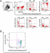
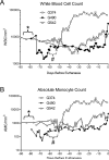
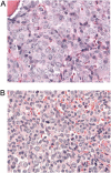
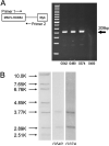
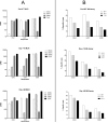
Results: Six weeks after infusion of HOXB4-overexpressing leukemia cells, these two dogs rapidly developed myeloid leukemia consisting of marrow and organ infiltration, circulating blasts, and, in one dog, chloromatous masses. Despite neither of these dogs sharing any dog leukocyte antigen haplotypes with the sentinel case, the HOXB4-transduced clones engrafted and proliferated without difficulty in the presence of immunosuppression. Chimerism studies in both dogs confirmed that donor and, in one case, host hematopoietic cell engraftment was lost and replaced by third-party HOXB4 cells.
Conclusions: The engraftment and expansion of these leukemia cells in dogs will allow studies into the biology of leukemia and development and evaluation of novel antileukemia therapies in a clinically relevant large animal model.
Figures



Similar articles
-
High incidence of leukemia in large animals after stem cell gene therapy with a HOXB4-expressing retroviral vector.J Clin Invest. 2008 Apr;118(4):1502-10. doi: 10.1172/JCI34371. J Clin Invest. 2008. PMID: 18357342 Free PMC article.
-
High-level ectopic HOXB4 expression confers a profound in vivo competitive growth advantage on human cord blood CD34+ cells, but impairs lymphomyeloid differentiation.Blood. 2003 Mar 1;101(5):1759-68. doi: 10.1182/blood-2002-03-0767. Epub 2002 Oct 24. Blood. 2003. PMID: 12406897
-
Effects of HOXB4 overexpression on ex vivo expansion and immortalization of hematopoietic cells from different species.Stem Cells. 2007 Aug;25(8):2074-81. doi: 10.1634/stemcells.2006-0742. Epub 2007 May 17. Stem Cells. 2007. PMID: 17510218
-
[Ex vivo expansion of human hematopoietic stem cells by passive transduction of the HOXB4 homeoprotein].J Soc Biol. 2006;200(3):235-41. doi: 10.1051/jbio:2006027. J Soc Biol. 2006. PMID: 17417138 Review. French.
-
Hox genes: from leukemia to hematopoietic stem cell expansion.Ann N Y Acad Sci. 2005 Jun;1044:109-16. doi: 10.1196/annals.1349.014. Ann N Y Acad Sci. 2005. PMID: 15958703 Review.
Cited by
-
Developments and translational relevance for the canine haematopoietic cell transplantation preclinical model.Vet Comp Oncol. 2020 Dec;18(4):471-483. doi: 10.1111/vco.12608. Epub 2020 May 26. Vet Comp Oncol. 2020. PMID: 32385957 Free PMC article. Review.
-
Insights into leukemia-initiating cell frequency and self-renewal from a novel canine model of leukemia.Exp Hematol. 2011 Jan;39(1):124-32. doi: 10.1016/j.exphem.2010.09.012. Epub 2010 Oct 8. Exp Hematol. 2011. PMID: 20933571 Free PMC article.
-
Animal Models for Preclinical Development of Allogeneic Hematopoietic Cell Transplantation.ILAR J. 2018 Dec 31;59(3):263-275. doi: 10.1093/ilar/ily006. ILAR J. 2018. PMID: 30010833 Free PMC article.
-
Future perspectives: therapeutic targeting of notch signalling may become a strategy in patients receiving stem cell transplantation for hematologic malignancies.Bone Marrow Res. 2011;2011:570796. doi: 10.1155/2011/570796. Epub 2010 Oct 4. Bone Marrow Res. 2011. PMID: 22046566 Free PMC article.
-
Evaluation of posttransplant methotrexate to facilitate engraftment in the canine major histocompatibility complex-haploidentical nonmyeloablative transplant model.Transplantation. 2010 Jul 15;90(1):14-22. doi: 10.1097/tp.0b013e3181e0a0c4. Transplantation. 2010. PMID: 20626083 Free PMC article.
References
-
- Fomchenko EI, Holland EC. Mouse models of brain tumors and their applications in preclinical trials (Review). Clin Cancer Res. 2006;12:5288–5297. - PubMed
-
- Felsburg PJ. Overview of immune system development in the dog: comparison with humans (Review). Human & Experimental Toxicology. 2002;21:487–492. - PubMed
-
- Joy F, Basak S, Gupta SK, Das PJ, Ghosh SK, Ghosh TC. Compositional correlations in canine genome reflects similarity with human genes. Journal of Biochemistry and Molecular Biology. 2006;39:240–246. - PubMed
-
- Deeg HJ, Storb R, Weiden PL, et al. Cyclosporin A and methotrexate in canine marrow transplantation: engraftment, graft-versus-host disease, and induction of tolerance. Transplantation. 1982;34:30–35. - PubMed
Publication types
MeSH terms
Substances
Grants and funding
LinkOut - more resources
Full Text Sources
Research Materials

