

Association of coronary aortic calcium with abdominal aortic calcium detected on lateral dual energy x-ray absorptiometry spine images
- PMID: 19616658
- PMCID: PMC2763778
- DOI: 10.1016/j.amjcard.2009.03.041
Association of coronary aortic calcium with abdominal aortic calcium detected on lateral dual energy x-ray absorptiometry spine images
Abstract
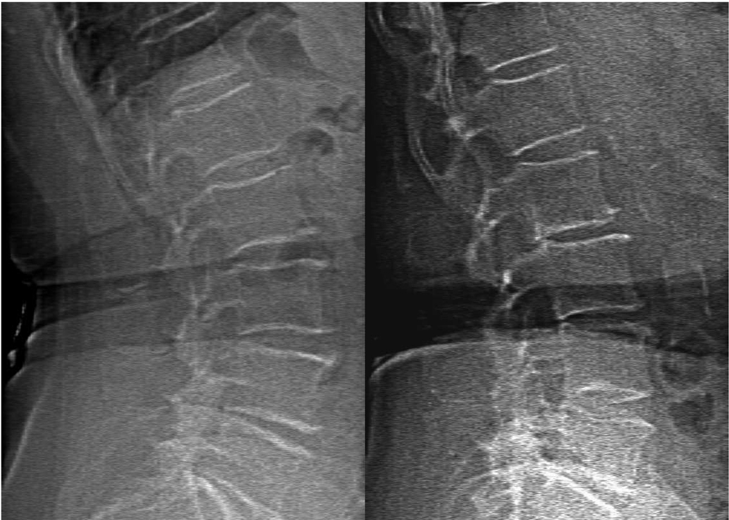
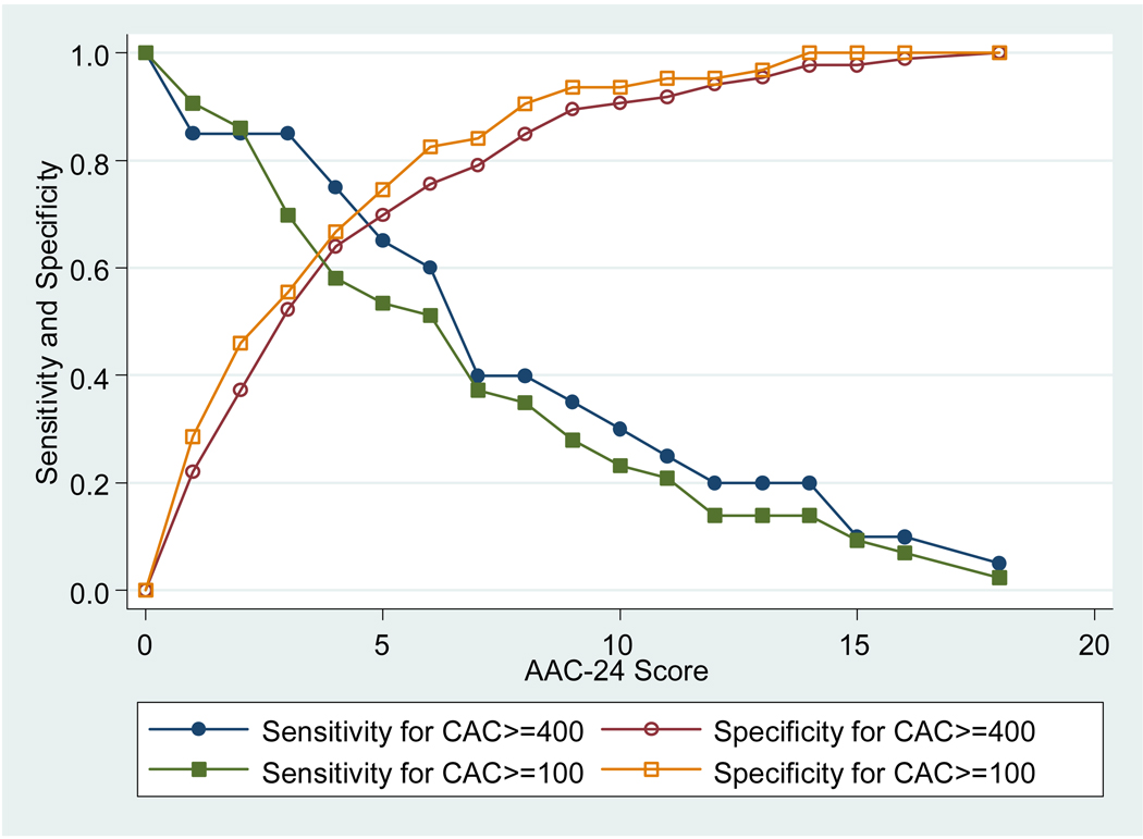
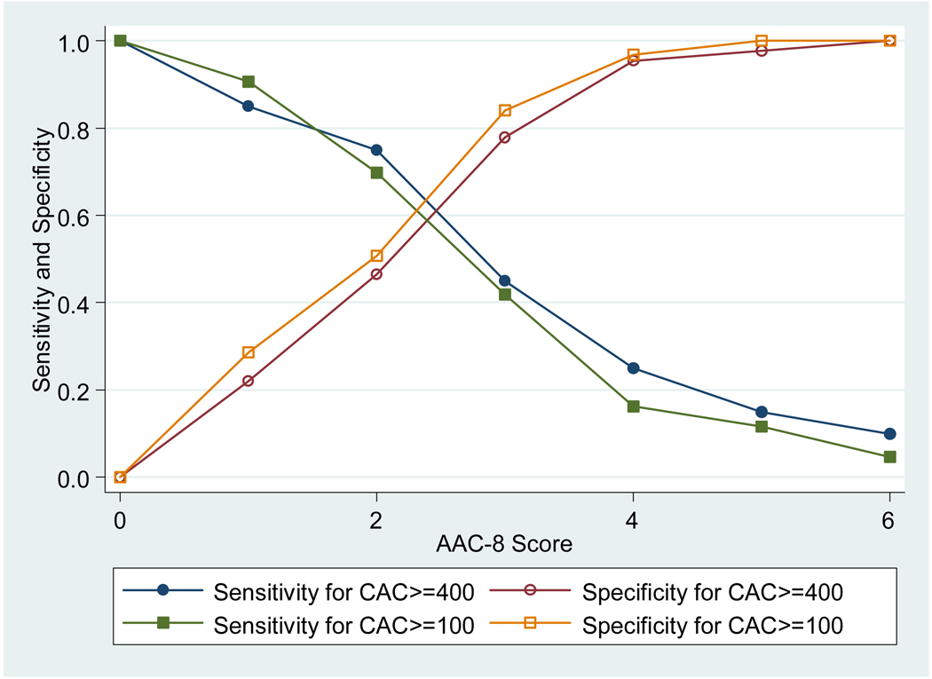
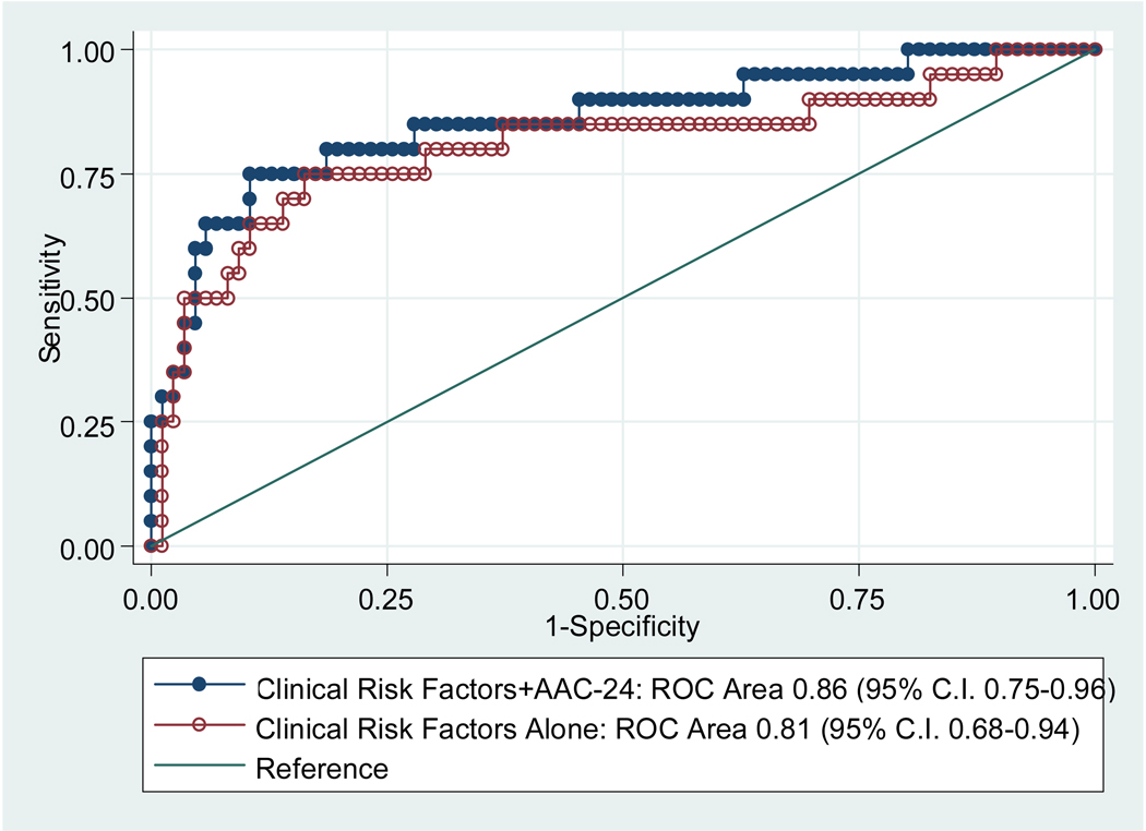
The association of abdominal aortic calcium (AAC) on lateral spine bone densitometry with coronary artery calcium (CAC) has not been reported. We studied 33 men and 73 women who had CAC scored with electron beam computed tomography at the eighth visit of the Rancho Bernardo study and lateral spine dual-energy x-ray absorptiometry images fully evaluable for AAC at the ninth study visit. The association between CAC level and AAC tertile was assessed by ordinal logistic regression analysis. The odds ratio of having a greater CAC score for those with an AAC score in the top tertile (24-point scale score > or =5) was 6.42 (95% confidence interval 2.28 to 18.1) and using the 8-point scale (score > or =3) was 3.38 (95% confidence interval 1.26 to 9.07) compared with those with AAC scores in the bottom tertiles, adjusted for age, gender, systolic blood pressure, total and high-density lipoprotein cholesterol, smoking status, and diabetes. A 24-point AAC score of > or =5 had a sensitivity of 65% and specificity of 70% to detect a high CAC score (> or =400 points). An 8-point AAC score of > or =3 had a sensitivity of 45% and specificity of 78%. In conclusion, a high level of AAC on lateral spine dual-energy x-ray absorptiometry was strongly associated with coronary artery disease and might be commonly encountered because bone densitometry is indicated for all women aged > or =65 years and all men aged > or =70 years. Its presence should be reported to the patient's physician to identify and manage modifiable risk factors.
Figures
References
-
- Schousboe JT, Taylor BC, Kiel DP, Ensrud KE, Wilson KE, McCloskey EV. Abdominal aortic calcification detected on lateral spine images from a bone densitometer predicts incident myocardial infarction or stroke in older women. J Bone Miner Res. 2008;23(3):409–416. - PubMed
-
- Agatston AS, Janowitz WR, Hildner FJ, Zusmer NR, Viamonte M, Jr, Detrano R. Quantification of coronary artery calcium using ultrafast computed tomography. J Am Coll Cardiol. 1990;15(4):827–832. - PubMed
-
- Kauppila LI, Polak JF, Cupples LA, Hannan MT, Kiel DP, Wilson PW. New indices to classify location, severity and progression of calcific lesions in the abdominal aorta: a 25-year follow-up study. Atherosclerosis. 1997;132(2):245–250. - PubMed
-
- Schousboe JT, Wilson KE, Kiel DP. Detection of abdominal aortic calcification with lateral spine imaging using DXA. J Clin Densitom. 2006;9(3):302–308. - PubMed
Publication types
MeSH terms
Grants and funding
LinkOut - more resources
Full Text Sources
Medical
