. 2009 Aug 1;74(5):1573-9.
doi: 10.1016/j.ijrobp.2009.04.017.

Interleukin-32 positively regulates radiation-induced vascular inflammation

Affiliations

Interleukin-32 positively regulates radiation-induced vascular inflammation

Hanako Kobayashi et al. Int J Radiat Oncol Biol Phys. .

Abstract

Purpose: To study the role of interleukin-32 (IL-32), a novel protein only detected in human tissues, in ionizing radiation (IR)-induced vascular inflammation.

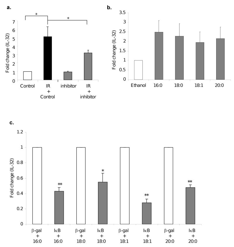
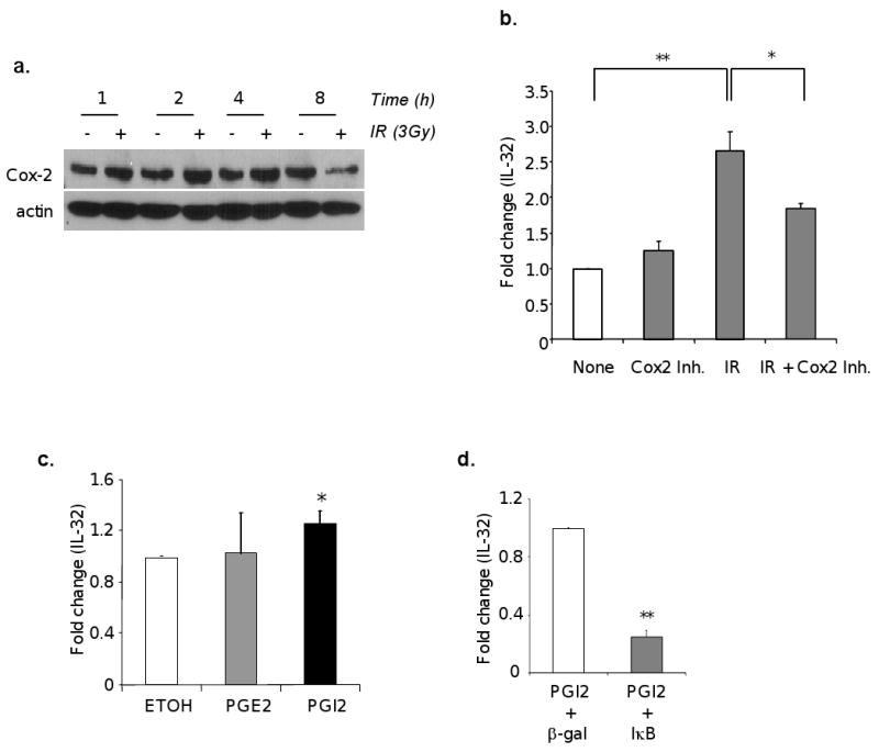
Methods and materials: Irradiated (0-6 Gy) human umbilical vein endothelial cells treated with or without various agents--a cytosolic phospholipase A2 (cPLA2) inhibitor, a cyclooxygenase-2 (Cox-2) inhibitor, or lysophosphatidylcholines (LPCs)--were used to assess IL-32 expression by Northern blot analysis and quantitative reverse transcriptase-polymerase chain reaction. Expression of cell adhesion molecules and leukocyte adhesion to endothelial cells using human acute monocytic leukemia cell line (THP-1) cells was also analyzed.

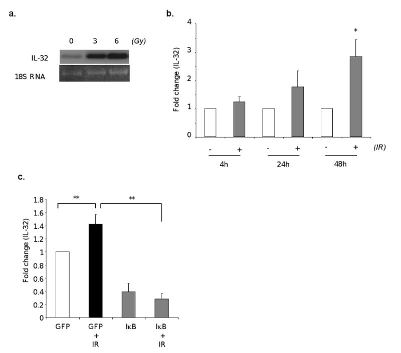
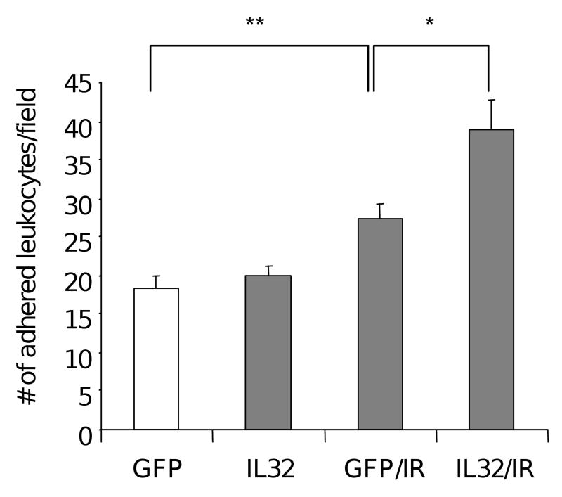
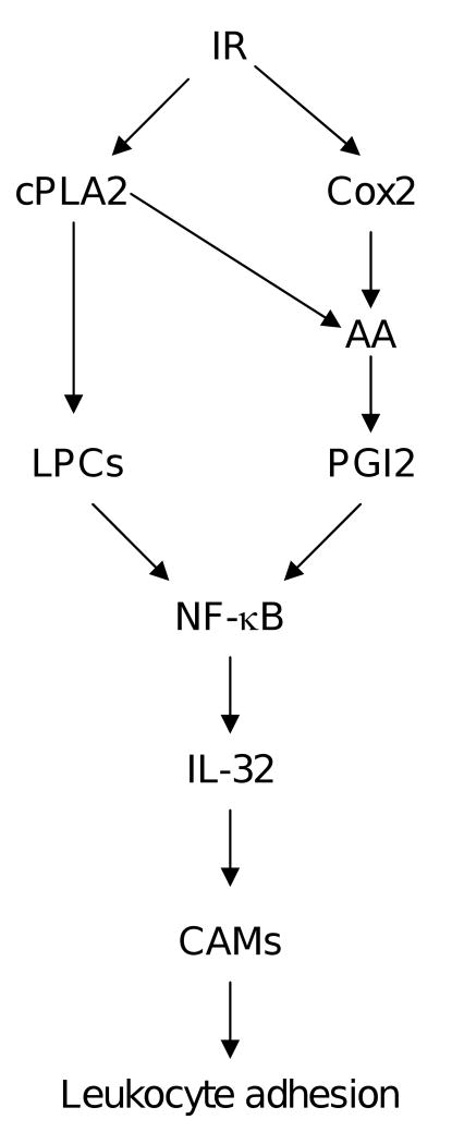
Results: Ionizing radiation dramatically increased IL-32 expression in vascular endothelial cells through multiple pathways. Ionizing radiation induced IL-32 expression through nuclear factor kappaB activation, through induction of cPLA2 and LPC, as well as induction of Cox-2 and subsequent conversion of arachidonic acid to prostacyclin. Conversely, blocking nuclear factor kappaB, cPLA2, and Cox-2 activity impaired IR-induced IL-32 expression. Importantly, IL-32 significantly enhanced IR-induced expression of vascular cell adhesion molecules and leukocyte adhesion on endothelial cells.

Conclusion: This study identifies IL-32 as a positive regulator in IR-induced vascular inflammation, and neutralization of IL-32 may be beneficial in protecting from IR-induced inflammation.


Conflict of interest statement

Conflict of Interest Notification: None.

Figures

Fig. 1
Fig. 1. IR induces IL-32 expression in human endothelial cells via NF-κB
HUVECs were irradiated at the indicated dose. Total RNAs were isolated 48 hours after irradiation and analyzed for IL-32 expression by Northern blot (a). HUVECs were irradiated with 0 or 3 Gy. Total RNAs were isolated at indicated time, and IL-32 levels were measured by qRT-PCR (b). HUVECs were infected with AdGFP or AdIκB at MOI 10. Following overnight infection, cells were irradiated at 0 or 3Gy, harvested 48 hours later. IL-32 levels were measured by qRT-PCR (c). * p<0.05, **p<0.01.
Fig. 2
Fig. 2. IL-32 induction by IR partly depends on cPLA2 that produces LPCs
HUVECs were treated with a Wyeth cPLA2 specific inhibitor at 10 μM or vehicle control for 30 min, followed by 3Gy IR. IL-32 levels were analyzed by qRT-PCR 48 hours after IR (a). *p<0.05. HUVECs were treated with indicated LPC species (20μM) at 0 and again at 24 hours. Total RNAs were isolated at 48 hours after IR and analyzed for IL-32 expression by qRT-PCR (b). HUVECs were infected with Adβ-gal or AdIκB at MOI=10. Following overnight infection, cells were treated with LPC for 48 hours. IL-32 levels were analyzed by qRT-PCR. Fold change of gene induction in the IκB group over the vector control was plotted (c). * p<0.05, **p<0.01.
Fig. 3
Fig. 3. IR upregulates IL-32 expression partly through induction of Cox2 and PGI2
HUVECs were treated with or without 3Gy IR. The cells were harvested at the time indicated after IR. Protein extract was subjected to Western immunoblot for Cox-2 levels (a). HUVECs were treated with vehicle control or celecoxib (25 uM) for 4 hours, and followed by IR exposure at 3 Gy. Total RNAs were isolated and IL-32 mRNA was measured by qRT-PCR (b). HUVECs were treated with PGE2 (10μM), PGI2 (10μM), or ethanol (a vehicle control) for 24 hours, and IL-32 mRNA was measured using qRT-PCR (c). HUVECs were infected with Adβ-gal or AdIκB at MOI=10. Following overnight infection, cells were treated with PGI2 for 24 hours, and IL-32 mRNA was measured using qRT-PCR (d). *p<0.05, **p<0.01.
Fig. 4
Fig. 4. Expression of IL-32 enhanced IR-induced leukocyte adhesion on vascular endothelium
Leukocyte cell adhesion was performed on irradiated (3Gy) HUVECs transfetced with a plasmid vector expressing either IL-32 or GFP control 48 hours after IR, respectively. Following a 30 min of incubation with dye labeled THP-1 cells, adhered cells were visualized and counted in high power fields under fluorescent microscopy. The experiments were performed with 6 wells per group, and repeated 3 times. *p<0.05, **p<0.01.
Fig. 5
Fig. 5. Schematic diagram illustrating the gene regulation mechanisms of IR-induced IL-32 gene expression, and its role in vascular inflammation

References

    1. Movsas B, Raffin TA, Epstein AH, et al. Pulmonary radiation injury. Chest. 1997;111:1061–1076. - PubMed
    1. Hallahan DE, Virudachalam S. Ionizing radiation mediates expression of cell adhesion molecules in distinct histological patterns within the lung. Cancer Res. 1997;57:2096–2099. - PubMed
    1. Hallahan DE, Virudachalam S, Kuchibhotla J. Nuclear factor kappaB dominant negative genetic constructs inhibit X-ray induction of cell adhesion molecules in the vascular endothelium. Cancer Res. 1998;58:5484–5488. - PubMed
    1. Kim SH, Han SY, Azam T, et al. Interleukin-32: a cytokine and inducer of TNFalpha. Immunity. 2005;22:131–142. - PubMed
    1. Shoda H, Fujio K, Yamaguchi Y, et al. Interactions between IL-32 and tumor necrosis factor alpha contribute to the exacerbation of immune-inflammatory diseases. Arthritis Res Ther. 2006;8:R166. - PMC - PubMed

Publication types

MeSH terms