. 2009 Jul 23;6(1):68-80.
doi: 10.1016/j.chom.2009.05.022.

A cellular restriction dictates the permissivity of nondividing monocytes/macrophages to lentivirus and gammaretrovirus infection

Affiliations

A cellular restriction dictates the permissivity of nondividing monocytes/macrophages to lentivirus and gammaretrovirus infection

Rajnish Kaushik et al. Cell Host Microbe. .

Abstract

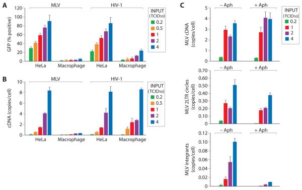
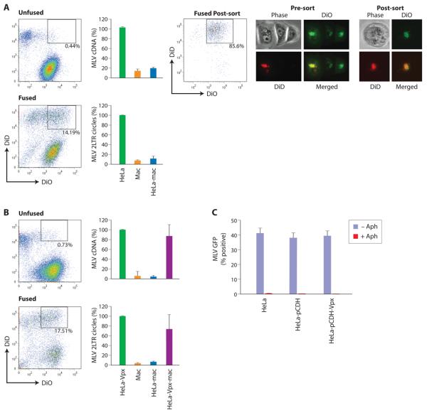
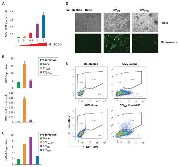
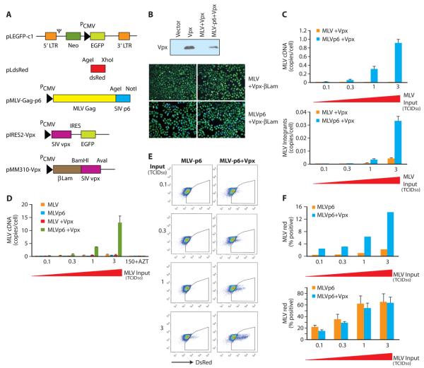
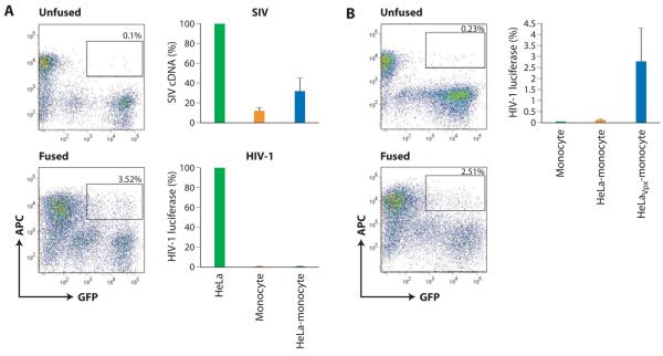
Primate lentiviruses, including HIV-1, transduce terminally differentiated, nondividing myeloid cells; however, these cells are refractory to infection by gammaretroviruses such as murine leukemia virus (MLV). Here, we present evidence that a cellular restriction is the obstacle to transduction of macrophages by MLV. Neutralization of the restriction by Vpx, a primate lentiviral protein previously shown to protect primate lentiviruses from a macrophage restriction, rendered macrophages permissive to MLV infection. We further demonstrate that this restriction prevents transduction of quiescent monocytes by HIV-1. Monocyte-HeLa heterokaryons were resistant to HIV-1 infection, while heterokaryons formed between monocytes and HeLa cells expressing Vpx were permissive to HIV-1 infection. Encapsidation of Vpx within HIV-1 virions conferred the ability to infect quiescent monocytes. Collectively, our results indicate that the relative ability of lentiviruses and gammaretroviruses to transduce nondividing myeloid cells is dependent upon their ability to neutralize a cellular restriction.


Figures

Figure 1
Figure 1. MLV infection of macrophage is blocked at, or prior, to reverse transcription of viral cDNA
Terminally differentiated macrophage and HeLa cells were infected with MLV and HIV-1 variants expressing GFP at different levels of input virions. The frequency of GFP positive cells (A) and viral cDNA copies (B) were determined 48 hours post infection. (C) MLV infection of aphidicolin treated and untreated HeLa cells. Viral cDNA (upper two panels) and viral integrants (lower panel) were determined at different levels of input virus based on tissue culture infectious dose50 (TCID50) where 1 TCID50 is the amount of virus inoculum that yielded 50% transduction on HeLa cells. (error bars are s.d. of replicate samples from 3 independent experiments done on macrophage from different donors).
Figure 2
Figure 2. A restriction prevents transduction of macrophage by MLV
Heterokaryons were formed between primary macrophage and between HeLa cells expressing fusogenic HN and F proteins of Newcastle Disease Virus (NDV). HeLa cells were stained with DiO (green) and macrophage were stained with DiD (red). Double-stained heterokaryons were sorted by FACS as indicated by the gate (A). FACS profile of heterokaryons post-sorting (fused post sort) is shown (middle panel) as are representative double staining heterokaryons pre-sort and post-sort (right panels). Because of the lipophilic nature of DiO and DiD, fluorescence concentrates in lipid-rich regions in the center of the cell rather than being evenly distributed throughout the cell. Susceptibility of HeLa-macrophage (HeLa-mac) heterokaryons to MLV infection was compared with infection levels in HeLa and in macrophage. Infection was gauged from the levels of late MLV cDNAs and 2-LTR circle cDNAs. Values were expressed relative to those obtained for HeLa cells (error bars are s.d. from 3 independent experiments). (B) Susceptibility of HeLa-macrophage heterokaryons to MLV infection was examined after expression of Vpx in HeLa cells. Double stained cells were sorted by FACS as indicated by the gate. MLV infection in HeLa-macrophage heterokaryons (HeLa-mac) and heterokaryons formed between macrophage and Vpx-expressing HeLa cells (HeLa-Vpx-mac) were gauged as outlined in A (error bars are s.d. of 3 independent experiments). (C) MLV infection of aphidicolin treated (+Aph) and untreated (−Aph) HeLa cells transfected with a Vpx expression vector (pCDH-Vpx) or an empty vector (pCDH).
Figure 3
Figure 3. Vpx permits transduction of macrophage by MLV in trans
(A) Vpx delivered to macrophage by wild-type SIV infection removes the block to synthesis of MLV cDNA in macrophage. Macrophage were initially infected with increasing titers of wild-type SIV and subsequently infected with MLV (4 TCID50) after 4 hours. Synthesis of MLV cDNA was assessed 48 hours after MLV infection. (B, C) Vpx but not Vpr is necessary for the ability of SIV to remove the block to macrophage transduction by MLV. Macrophage were infected by wild-type or Vpx-deleted SIV and subsequently infected by MLVGFP (4 TCID50) after 4 hours. In C, macrophages were infected with the indicated SIV infectious clones and then with MLV. The efficiency of MLV transduction was assessed after MLV infection. A representative field of macrophage transduced by MLV-GFP is shown in D. (E) Transduction of macrophage by MLV occurs primarily in SIV-infected macrophage. SIVGFP-infected macrophage were transduced with MLVdsRed and frequencies of coinfected cells was evaluated by FACS. FACS profiles of uninfected macrophage, MLV-transduced macrophage without prior SIV infection (MLV alone) or SIVWT without subsequent MLV infection (SIVWT alone) served as controls.
Figure 4
Figure 4. MLV virions encapsidating Vpx exhibit a lentiviral phenotype
(A) A schematic of vectors used for expression of Vpx and chimeric MLV gag proteins containing the p6 domain of SIV gag which harbors the Vpx/Vpr packaging determinant. (B) Packaging of Vpx within MLV virions harboring an SIV gag p6 domain. Upper panel, packaging of Vpx within MLV virions containing or lacking an SIV p6 gag domain was examined by Western blotting with a Vpx-specific antibody. Lower panels, Vpx-β-lactamase fusion proteins were packaged in MLV variants containing or lacking the SIV gag p6 domain and β-lactamase activity was examined following infection of HeLa cells loaded with the β-lactamase substrate CCF2. (C) Packaging of Vpx within chimeric MLV virions containing SIV gag p6 (MLVp6) removes a block to reverse transcription in macrophage. Macrophage were infected with increasing concentrations of MLVp6 with or without encapsidated Vpx and viral cDNA synthesis (late cDNA, upper panel) and integration (lower panel) was assessed. (D-F) A p6 encapsidation signal and Vpx is required for MLV transduction of macrophage. MLV cDNA synthesis (D) was examined after infection of macrophage with MLV and MLVp6 variants with and without Vpx. Infections carried out in the presence of AZT verified de novo synthesis of MLV cDNA, (E). Packaging of Vpx permits transduction of primary macrophage by MLV. Macrophage were infected with increasing titers of chimeric MLV variants with and without Vpx as in C. Transduction was gauged by expression of dsRed from the MLV transgene. Frequencies of MLV transduction (dsRed expression) on macrophage (upper panel) and HeLa (lower panel) are indicated in panel F.
Figure 5
Figure 5. Transduction of primary monocytes by HIV-1 is blocked by a restriction
Heterokaryons were formed between primary monocytes and HeLa cells using HVJ envelope fusion kit. (A) FACS analysis of HeLa-monocyte heterokaryons (left panels). HeLa cells expressed GFP and macrophages were stained with an APC-conjugated antibody to CD14. Double-stained cells were sorted as indicated by the gate. SIV infection was gauged from the levels of late cDNAs and HIV-1 infection was gauged from luciferase activity (right panels). Values were expressed relative to those obtained for HeLa cells (error bars are s.d. of 4 independent experiments.) (B) Vpx renders HeLa-monocyte heterokaryons permissive to HIV-1 infection. Heterokaryons were formed between primary monocytes and HeLa cells expressing Vpx as described in A). Susceptibility of HeLa-monocyte heterokaryons to HIV-1 infection was examined after expression of Vpx in HeLa cells. FACS analysis of HeLa-Vpx-monocyte heterokaryons is shown on the left panels. Double-stained cells were sorted as indicated by the gate. Infection of monocytes and infection of HeLa-monocyte heterokaryons with and without Vpx was gauged by luciferase activity (error bars are s.d. from 2 independent experiments).
Figure 6
Figure 6. Vpx counteracts a monocyte restriction to HIV-1 infection in trans
(A) Infection of monocytes by wild-type SIV removes a reverse transcription block to subsequent infection by HIV-1. SIVWT-infected monocytes were subsequently infected (4 hours later) by HIV-1 on the indicated intervals and levels of HIV-1 cDNA synthesis was gauged 48 hours post HIV-1 infection (B) Prior infection by wild-type but not Vpx-deleted SIV renders primary monocytes permissive to subsequent transduction by HIV-1. Monocytes were infected as in A. Transduction of HIV-1 (based on GFP expression) was assessed 72 hours post HIV-1 infection. Representative fields of primary monocytes following transduction by HIV-1-GFP are shown on C. (D) HIV-1 virions encapsidating Vpx efficiently transduce primary monocytes. Monocytes were infected with HIV-1 GFP variants in which Vpx was packaged. Levels of transduction (% GFP-positive monocytes) was determined at the indicated intervals post monocyte infection. (E) Transduction of monocytes with an HIV-1 lentivirus vector in which Vpx was or was not packaged. Monocytes were infected at the indicated intervals and GFP expression examined 72h post-infection (error bars are s.d. of replicate samples from 3 independent experiments done on macrophage from different donors).
Figure 7
Figure 7. Vpx renders monocytes permissive to HIV-1 infection without inducing monocyte differentiation or Apobec 3G distribution
(A) Distribution of Apobec 3G between LMM and HMM nucleoprotein complexes in undifferentiated (d0) monocytes, differentiated (d10) macrophage and SIV-infected monocytes. Distribution of Apobec 3G between H9 cell-derived HMM and LMM complexes before and after RNase treatment is shown for comparison. (B) Vpx does not affect differentiation status of monocytes in culture. Fresh monocytes were infected with HIV-1ΔVprGFP that had or had not packaged Vpx and the infection levels in monocyte/macrophage (CD14+) and differentiated monocyte (CD71+) subsets was determined at the indicated intervals post infection by FACS. (C) HIV-1 with encapsidated Vpx equally transduces undifferentiated (CD71) and differentiated (CD71+) monocyte populations. Monocytes were infected with HIV-1 in which Vpx had been packaged (lower three panels) and the frequencies of infected (GFP positive) CD71+ macrophage and CD71 monocytes were determined by FACS. Upper three panels depict uninfected controls. The frequency of HIV-1 infection in CD71+ and CD71 cells at different intervals post- infection is shown in D. Supporting Information

Similar articles

Cited by

References

    1. Accola MA, Bukovsky AA, Jones MS, Gottlinger HG. A conserved dileucine-containing motif in p6(gag) governs the particle association of Vpx and Vpr of simian immunodeficiency viruses SIV(mac) and SIV(agm) J Virol. 1999;73:9992–9999. - PMC - PubMed
    1. Alexaki A, Liu Y, Wigdahl B. Cellular reservoirs of HIV-1 and their role in viral persistence. Curr HIV Res. 2008;6:388–400. - PMC - PubMed
    1. Andrejeva J, Poole E, Young DF, Goodbourn S, Randall RE. The p127 subunit (DDB1) of the UV-DNA damage repair binding protein is essential for the targeted degradation of STAT1 by the V protein of the paramyxovirus simian virus 5. J Virol. 2002;76:11379–11386. - PMC - PubMed
    1. Arthur LO, Bess JW, Snowder RC, II, Benveniste RE, Mann DL, Chermann J-C, Henderson LE. Cellular proteins bound to immunodeficiency viruses: implications for pathogenesis and vaccines. Science. 1992;258:1935–1938. - PubMed
    1. Balcaitis S, Weinstein JR, Li S, Chamberlain JS, Moller T. Lentiviral transduction of microglial cells. Glia. 2005;50:48–55. - PubMed

Publication types

Substances

LinkOut - more resources