

doi: 10.1177/1099800409338516.
Epub 2009 Jul 17.
Basic concepts of optical mapping techniques in cardiac electrophysiology
Affiliations
- PMID: 19617237
- PMCID: PMC3167575
- DOI: 10.1177/1099800409338516
Item in Clipboard
Basic concepts of optical mapping techniques in cardiac electrophysiology
Biol Res Nurs.
2009 Oct.
Abstract
Optical mapping is a tool used in cardiac electrophysiology to study the heart's normal rhythm and arrhythmias. The optical mapping technique provides a unique opportunity to obtain membrane potential recordings with a higher temporal and spatial resolution than electrical mapping. Additionally, it allows simultaneous recording of membrane potential and calcium transients in the whole heart. This article presents the basic concepts of optical mapping techniques as an introduction for students and investigators in experimental laboratories unfamiliar with it.
Figures
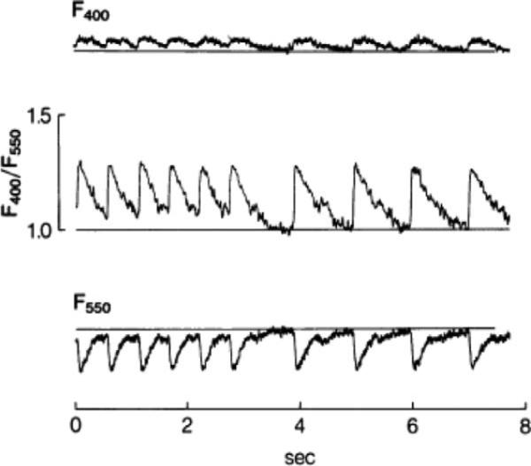
First recording of calcium transients in intact heart loaded with Indo-1. Fluorescence increases at 400 ± 12.5 nm (top trace) and decreases at 550 ± 20 nm (bottom trace). In the middle trace, the first six beats of calcium transients were induced by pacing of right ventricle whereas the last four beats occurred spontaneously. The fluorescence was collected by fiber-optic cables before reaching photomultiplier tubes. The photomultiplier is a photodetector used when the light level is very low (Adapted from Lee et al., 1987). Reprinted with permission.
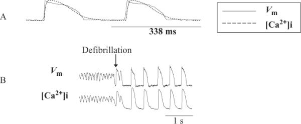
Simultaneous recording of membrane potential and calcium transients during normal sinus rhythm, ventricular fibrillation (VF), and defibrillation. (A) Optical signals from membrane potential and calcium transients during normal sinus rhythm. (B) Optical signals during VF, defibrillation and normal sinus rhythm in Langendorff-perfused rabbit heart (Attin, 2005).
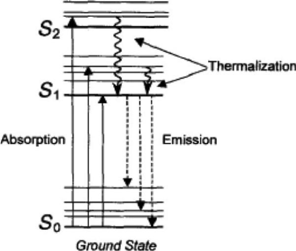
Perrin-Jablonski diagram. S0 = ground state; S1 = excited state (Adapted from Jameson et al., 2003). Reprinted with permission.
References
-
- Allessie MA, Bonke FI, Schopman FJ. Circus movement in rabbit atrial muscle as a mechanism of tachycardia. III. The “leading circle” concept: A new model of circus movement in cardiac tissue without the involvement of an anatomical obstacle. Circulation Research. 1977;41:9–18. - PubMed
-
- Attin M. Unpublished doctoral dissertation. University of California; Los Angeles: 2005. Calcium transients: An insight into ventricular defibrillation.
-
- Baker LC, Wolk R, Choi B-R, Watkins S, Plan P, Shah A, et al. Effects of mechanical uncouplers, diacetyl monoxime, and cytochalasin-D on the electro-physiology of perfused mouse hearts. American Journal of Physiology. 2004;287:H1771–H1779. - PubMed
-
- Banville I, Gray R. Effect of action potential duration and conduction velocity restitution and their spatial dispersion on alternans and the stability of arrhythmias. Journal of Cardiovascular Electrophysiology. 2002;13:1141–1149. - PubMed
-
- Baxter WT, Davidenko JM, Loew LM, Wuskell JP, Jalife J. Technical features of a CCD video camera system to record cardiac fluorescence data. Annals of Biomedical Engineering. 1997;25:713–725. - PubMed
Publication types
MeSH terms
Grants and funding
LinkOut - more resources
Full Text Sources
Medical
