

Staging of cutaneous melanoma
- PMID: 19617293
- PMCID: PMC2712594
- DOI: 10.1093/annonc/mdp256
Staging of cutaneous melanoma
Abstract
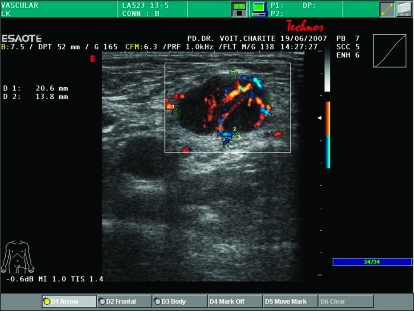
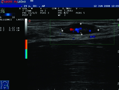
The American Joint Committee on Cancer (AJCC) staging of cutaneous melanoma is a continuously evolving system. The identification of increasingly more accurate prognostic factors has led to major changes in melanoma staging over the years, and the current system described in this review will likely be modified in the near future. Likewise, application of new imaging techniques has also changed the staging work-up of patients with cutaneous melanoma. Chest and abdominal computed tomography (CT) scanning is most commonly used for evaluation of potential metastatic sites in the lungs, lymph nodes and liver, and is indicated in patients with new symptoms, anaemia, elevated lactate dehydrogenase or a chest X-ray abnormality. CT scans should be restricted to patients with high-risk melanoma (stage IIC, IIIB, IIIC and stage IIIA with a macroscopic sentinel lymph node). Magnetic resonance imaging (MRI) of the brain is a mandatory test in patients with stage IV, optional in stage III and not used in patients with stage I and II disease. Positron emission tomography (PET)/CT is more accurate than CT or MRI alone in the diagnosis of metastases and should complement conventional CT/MRI imaging in the staging work-up of patients who have solitary or oligometastatic disease where surgical resection is most relevant.
Figures
References
-
- Balch CM, Buzaid AC, Soong SJ, et al. Final version of the American Joint Committee on Cancer staging system for cutaneous melanoma. J Natl Compr Canc Netw. 2006;4:666–684.
-
- Garbe C, Hauschild A, Volkenandt M, et al. Evidence and interdisciplinary consense-based German guidelines: diagnosis and surveillance of melanoma. Melanoma Res. 2007;17:393–399. - PubMed
-
- Dummer R, Panizzon R, Bloch PH, Burg G. Updated Swiss guidelines for the treatment and follow-up of cutaneous melanoma. Dermatology. 2005;210:39–44. - PubMed
-
- Davis SD. CT evaluation for pulmonary metastases in patients with extrathoracic malignancy. Radiology. 1991;180:1–12. - PubMed
-
- Heaston DK, Putman CE, Rodan BA, et al. Solitary pulmonary metastases in high-risk melanoma patients: a prospective comparison of conventional and computed tomography. AJR Am J Roentgenol. 1983;141:169–174. - PubMed
Publication types
MeSH terms
LinkOut - more resources
Full Text Sources
Medical
