. 2009 Jul 28;106(30):12329-34.
doi: 10.1073/pnas.0904175106. Epub 2009 Jul 15.

Energetics of glutamate receptor ligand binding domain dimer assembly are modulated by allosteric ions

Affiliations

Energetics of glutamate receptor ligand binding domain dimer assembly are modulated by allosteric ions

Charu Chaudhry et al. Proc Natl Acad Sci U S A. .

Abstract

The activity of many ligand-gated ion channels and cell surface receptors is modulated by small molecules and ions, but an understanding of the underlying molecular mechanisms is scarce. For kainate, but not AMPA subtype glutamate receptors, the binding of Na(+) and Cl(-) ions to discrete, electrostatically coupled sites in the extracellular ligand binding domain (LBD) dimer assembly regulates the rate of entry into the desensitized state, which occurs when the dimer interface ruptures and the channel closes. Studies on glutamate receptors have defined the LBD dimer assembly as a key functional unit that controls activation and desensitization. Here we use analytical ultracentrifugation to probe the energetic effects of allosteric ions on kainate receptor dimer stability in solution, using a GluR6 mutant that desensitizes slowly. Our results show that sodium and chloride ions modulate kainate receptor dimer affinity as much as 50-fold, and that removal of either Cl(-) or Na(+) disrupts the dimer. The applicability of a similar allosteric mechanism for modulation of delta2 glutamate receptors by Ca(2+) was also tested. Our results indicate that ions can contribute substantial free energy to active state stabilization in both these receptors, and provide quantitative measurements of the energetic consequences of allosteric ion binding to a ligand-gated ion channel.


Conflict of interest statement

The authors declare no conflict of interest.

Figures
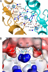
Fig. 1.
Fig. 1.
The Na+ and Cl binding sites in GluR6 HERLK. (A) Crystal structure of the 1.3 Å resolution glutamate complex (PDB 3G3J) looking down the molecular 2-fold onto the membrane plane; the 2 subunits of the dimer are colored gold and cyan. σ A-weighted 2mFo-DFc electron density maps contoured at 2σ are shown for Na+ (yellow spheres) and Cl (green sphere) ions. (B) Electrostatic potential map of the protein interior contoured from −10kT/e (red) to +10kT/e (blue); view is rotated ≈90° from that in A. Side chains and solvent forming the pentavalent Na+ and octahedral Cl coordination spheres are shown; the electrostatic potential map was calculated for the high occupancy conformation of R744.
Fig. 2.
Fig. 2.
Ion sensitivity of GluR6 HERLK resembles that for wt. (A) The kinetics of entry into desensitization are slowed for both wt GluR6 and HERLK when NaCl is increased from 150 to 600 mM. The glutamate application was 100 ms for wt and 7 s for HERLK. (B) Modulation of HERLK currents by cations and anions; the current is almost entirely abolished in CsMeSO3. (C) The desensitization of HERLK currents is well described by 2 exponential decays for a range of ionic conditions. (D) Rates of entry into desensitization for wild-type and HERLK mutant receptors show a linear correlation across the entire range of ionic conditions tested (R = 0.99).
Fig. 3.
Fig. 3.
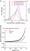
Na+ and Cl ions modulate LBD dimer stability. (A) Sedimentation velocity c(s) distributions for GluR6 HERLK in 165 and 600 mM NaCl (blue and red traces) and 165 mM CsMeSO3 (black traces); values in brackets indicate protein concentrations calculated from integration of the c(s) distributions. In CsMeSO3, the protein is monomeric (2.6S). 600 mM NaCl, the profile, reflecting a rapidly interconverting monomer-dimer system, shifts to sw values of NB approximately 3.5 and 3.7S at loading concentrations of 0.3 and 2 mg/mL (dashed and solid red lines, respectively), approaching the expected 4S value for a dimer. (B) Representative sedimentation equilibrium distribution of HERLK in 600 mM NaCl at 24,000 rpm. Global fits to a monomer-dimer model yields a Kd of 12 μM (95% confidence interval of 11–13 μM). (Upper) Circles are the data points, the red line is the global fit, and dotted and dashed lines are the exponentials representing monomer and dimer fractions whose sum gives rise to this fit; (Lower) residuals are shown.
Fig. 4.
Fig. 4.
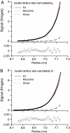
Dimer assembly is disrupted on removal of either anion or cation. Representative sedimentation equilibrium profiles for GluR6 HERLK at 24,000 rpm with Cl replaced by MeSO3 (A) and Na+ by NMDG+ (B). The dimerization Kd decreases about 50-fold in both cases yielding values of 610 μM (95% confidence interval of 450–890 μM) for NaMeSO3 and 550 μM (510–600 μM) for NMDG-Cl.
Fig. 5.
Fig. 5.
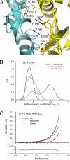
Ca2+ stabilizes the active dimer assembly in GluR δ2. (A) Top view of the GluRδ2-S1S2 dimer structure (PDB 2V3T) showing 2 bound Ca2+ ions (gold spheres) forming pentavalent coordination complexes that link the dimer assembly (16, 23). Side chains for R781 and T538, equivalent to R744 and K500 in GluR6, are shown to highlight the absent anion site. (B) Sedimentation coefficient distributions of HYLAK δ2 yields Sw values of 3.42S in 20 mM CaCl2 (2.4 mg/mL), 2.76S in 20 mM MgCl2 (2.5 mg/mL), and 2.69S in 2 mM EGTA (2.4 mg/mL); protein concentrations are derived from integrated peaks in c(s) distributions. (C) Representative sedimentation equilibrium distribution of HYLAKδ2 in 20 mM CaCl2 derived from a global analysis of data at 3 concentrations and 3 rotor speeds, using a monomer-dimer model with a Kd of 62 μM (95% confidence interval of 51–74 μM). Graph shows data points at 24,000 rpm, the black line is the model used to fit the data, and dotted and dashed lines represent the best-fit populations of monomer and dimer respectively; residuals of the fit are shown below the graph.

References

    1. Changeux JP, Edelstein SJ. Allosteric receptors after 30 years. Neuron. 1998;21:959–980. - PubMed
    1. Sun Y, et al. Mechanism of glutamate receptor desensitization. Nature. 2002;417:245–253. - PubMed
    1. Jin R, et al. Mechanism of positive allosteric modulators acting on AMPA receptors. J Neurosci. 2005;25:9027–9036. - PMC - PubMed
    1. Silberberg SD, Li M, Swartz KJ. Ivermectin interaction with transmembrane helices reveals widespread rearrangements during opening of P2X receptor channels. Neuron. 2007;54:263–274. - PubMed
    1. Ymer S, et al. Structural and functional characterization of the gamma 1 subunit of GABAA/benzodiazepine receptors. EMBO J. 1990;9:3261–3267. - PMC - PubMed

Publication types

MeSH terms

LinkOut - more resources