. 2009 Aug 4;106(31):12776-81.
doi: 10.1073/pnas.0903075106. Epub 2009 Jul 17.

Heterozygosity for a Bub1 mutation causes female-specific germ cell aneuploidy in mice

Affiliations

Heterozygosity for a Bub1 mutation causes female-specific germ cell aneuploidy in mice

Shawn Leland et al. Proc Natl Acad Sci U S A. .

Abstract

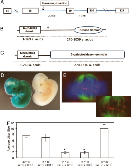
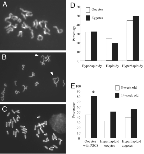
Aneuploidy, the most common chromosomal abnormality at birth and the main ascertained cause of pregnancy loss in humans, originates primarily from chromosome segregation errors during oogenesis. Here, we report that heterozygosity for a mutation in the mitotic checkpoint kinase gene, Bub1, induces aneuploidy in female germ cells of mice and that the effect increases with advancing maternal age. Analysis of Bub1 heterozygous oocytes showed that aneuploidy occurred primarily during the first meiotic division and involved premature sister chromatid separation. Furthermore, aneuploidy was inherited in zygotes and resulted in the loss of embryos after implantation. The incidence of aneuploidy in zygotes was sufficient to explain the reduced litter size in matings with Bub1 heterozygous females. No effects were seen in germ cells from heterozygous males. These findings show that Bub1 dysfunction is linked to inherited aneuploidy in female germ cells and may contribute to the maternal age-related increase in aneuploidy and pregnancy loss.


Conflict of interest statement

The authors declare no conflict of interest.

Figures

Fig. 1.
Fig. 1.
Bub1 mutant females are subfertile. (A) Gene structure of the murine Bub1. The relative insertion site is indicated by an arrow. (B) Bub1 protein organization. The functional domains within the Bub1 protein are shown, and the relative trap insertion site is indicated by an arrow. (C) Bub1-βgeo mutant protein organization. The replacement of the C-terminal amino acids by the β-galactosidase-neomycin (β-geo) fusion peptide is shown. (D) Expression of Bub1 in 10.5 dpc embryos using the β-geo reporter mediated conversion of X-gal substrate. Photograph shows a Bub1 heterozygote embryo (stained blue) and a control WT littermate embryo obtained from a WT female mouse crossed to a Bub+/m male. (E) Localization of the mutant Bub1-β-geo fusion peptide to kinetochores in Bub+/m mouse embryonic fibroblasts (MEFs). Immunofluorescence analysis of a metaphase cell stained with FITC-conjugated α-tubulin antibodies to visualize microtubules (green), anti-β-galactosidase antibodies bound to Texas-red conjugated secondary antibodies (red), and DAPI stained as blue to visualize DNA is shown above. Note the dotted appearance of the Bub1-β-geo fusion protein signals at the chromosome-microtubule junctions that indicate kinetochore localization. Inset shows the same image without the DAPI-stained DNA for better visualization of the kinetochore localization of the Bub1-β-galactosidase fusion protein. (F) The average litter size of various intercrosses obtained from Bub1+/m and WT mice are shown. The bars indicate the standard error within the dataset obtained from each intercross and n refers to the total number of crosses from each intercross. The differences between the litter sizes obtained from the heterozygous females were significantly different from the intercrosses obtained from the WT females. ∗, P < 0.0001, Student t test.
Fig. 2.
Fig. 2.
Metaphase II oocytes and zygotes from Bub1+/m females have abnormal chromosome numbers. Bub1+/m or 8- to 16-week-old WT females were mated with B6C3F1 males and zygotes collected ≈30 h after the induction of superovulation. Oocytes were collected from females that did not mate ≈20 h after the induction of superovulation. (A) Normal metaphase II oocyte with 20 dyads from a WT female. (B) Metaphase II oocyte from a Bub1+/m female with a total of 21 chromosomes. Arrowheads indicate single chromatids. (C) Zygote from a Bub+/m female with 20 paternal chromosomes on the left and 23 maternal chromosomes on the right. (D) Comparisons of aneuploidy frequencies in metaphase II oocytes and zygotes from Bub+/m females showing similar levels of hypohaploid and hyperhaploid maternal complements before and after fertilization. (E) Age-dependent increase in the incidences of PSCS and hyperploid oocytes and zygotes in Bub1+/m female of different ages. ∗, P < 0.002 (χ2).

References

    1. Hassold T, Hall H, Hunt P. The origin of human aneuploidy: Where we have been, where we are going. Hum Mol Genet. 2007;16(Spec No 2):R203–R208. - PubMed
    1. Hassold T, Hunt P. To err (meiotically) is human: The genesis of human aneuploidy. Nat Rev Genet. 2001;2:280–291. - PubMed
    1. Vogt E, Kirsch-Volders M, Parry J, Eichenlaub-Ritter U. Spindle formation, chromosome segregation and the spindle checkpoint in mammalian oocytes and susceptibility to meiotic error. Mutat Res. 200;651:14–29. - PubMed
    1. Musacchio A, Salmon ED. The spindle-assembly checkpoint in space and time. Nat Rev Mol Cell Biol. 2007;8:379–393. - PubMed
    1. Amon A. The spindle checkpoint. Curr Opin Genet Dev. 1999;9:69–75. - PubMed

Publication types