. 2009 Oct 16;284(42):28616-23.
doi: 10.1074/jbc.M109.017525. Epub 2009 Jul 18.

Cell adhesion to tropoelastin is mediated via the C-terminal GRKRK motif and integrin alphaVbeta3

Affiliations

Cell adhesion to tropoelastin is mediated via the C-terminal GRKRK motif and integrin alphaVbeta3

Daniel V Bax et al. J Biol Chem. .

Abstract

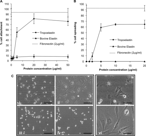
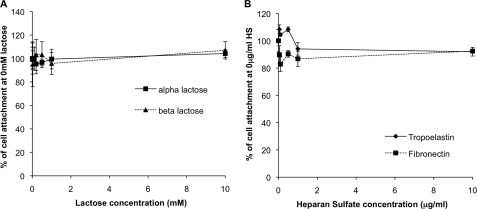
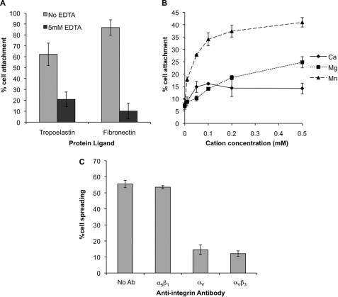
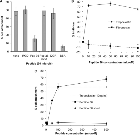
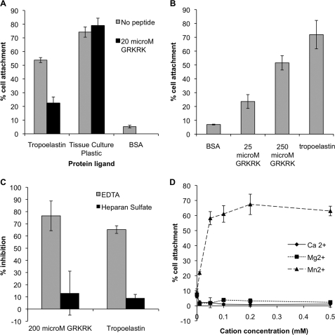
Elastin fibers are predominantly composed of the secreted monomer tropoelastin. This protein assembly confers elasticity to all vertebrate elastic tissues including arteries, lung, skin, vocal folds, and elastic cartilage. In this study we examined the mechanism of cell interactions with recombinant human tropoelastin. Cell adhesion to human tropoelastin was divalent cation-dependent, and the inhibitory anti-integrin alpha(V)beta(3) antibody LM609 inhibited cell spreading on tropoelastin, identifying integrin alpha(V)beta(3) as the major fibroblast cell surface receptor for human tropoelastin. Cell adhesion was unaffected by lactose and heparin sulfate, indicating that the elastin-binding protein and cell surface glycosaminoglycans are not involved. The C-terminal GRKRK motif of tropoelastin can bind to cells in a divalent cation-dependent manner, identifying this as an integrin binding motif required for cell adhesion.


Figures

FIGURE 1.
FIGURE 1.
A and B, human dermal fibroblast attachment (A) and spreading (B) to a concentration gradient of recombinant human tropoelastin (diamonds, solid line) and bovine soluble elastin (triangles, solid line). The dashed line indicates the % cell attachment or spreading onto the positive control, 2 μg/ml human plasma fibronectin. C, phase contrast photographs of human dermal fibroblast spreading onto 5 μg/ml recombinant human tropoelastin (i and v), 2 μg/ml human plasma fibronectin (ii and vi), 5 μg/ml bovine soluble elastin (iii), or BSA-blocked wells (iv) after incubation for 90 min. Scale bars on i–iv indicate 200 μm and on v and vi indicate 100 μm. All error bars indicate S.D. of triplicate measurements.
FIGURE 2.
FIGURE 2.
A, inhibition of human dermal fibroblast attachment onto 10 μg/ml recombinant human tropoelastin by α lactose (squares, solid line) or β lactose (triangles, dashed line). B, inhibition of human dermal fibroblast attachment onto 10 μg/ml recombinant human tropoelastin (diamonds, solid line) or 2 μg/ml human plasma fibronectin (squares, dashed line) using a concentration gradient of heparan sulfate (HS). Error bars indicate S.D. of triplicate measurements.
FIGURE 3.
FIGURE 3.
A, inhibition of human dermal fibroblast attachment to 10 μg/ml recombinant human tropoelastin or 2 μg/ml human plasma fibronectin in the presence (dark gray bars) or absence (light gray bars) of 5 mm EDTA. B, human dermal fibroblast attachment on to 10 μg/ml recombinant human tropoelastin in the presence of a concentration gradient of Ca2+ (diamonds), Mg2+ (squares), or Mn2+ (triangles). In additional studies, the degree of attachment in the presence of 0.5 mm Mn2+ was similar to that in Dulbecco's modified Eagle's medium, 49.5% for 0.5 mm Mn2+ and 48% for Dulbecco's modified Eagle's medium (data not shown). C, anti-integrin antibody (Ab) inhibition of human dermal fibroblast spreading onto 10 μg/ml recombinant human tropoelastin. Antibodies JBS-5 (anti α5β1), 17E6 (anti αV), and LM609 (anti αVβ3) were used at 20 μg/ml. Error bars indicate S.D. of triplicate measurements.
FIGURE 4.
FIGURE 4.
A, inhibition of human dermal fibroblast attachment to 10 μg/ml human recombinant tropoelastin with 50 μm RGD peptide, Peptide 36 (ACLGKACGRKRK), Peptide 36 Short (ACLGKACG), and the negative control DGR. B, dose-dependent inhibition of human dermal fibroblast attachment to 10 μg/ml human recombinant tropoelastin (diamonds, solid line) or 1 μg/ml human plasma fibronectin (squares, dashed line) with a concentration gradient of Peptide 36. Percentage inhibitions were calculated against cell adhesion to a non-blocked control. C, human dermal fibroblast attachment to wells coated with a concentration gradient of Peptide 36 (squares) or Peptide 36 Short (triangles). The percentage of cell attachment to wells coated with 10 μg/ml human recombinant tropoelastin is indicated with a dashed line. Error bars indicate S.D. of triplicate measurements.
FIGURE 5.
FIGURE 5.
A, human dermal fibroblast attachment to wells coated with 10 μg/ml human recombinant tropoelastin then BSA blocked or to uncoated and unblocked tissue culture wells in the presence (black bars) or absence (gray bars) of 20 μm peptide GRKRK. B, human dermal fibroblast attachment to wells coated with 25 or 250 μm peptide GRKRK or 10 μg/ml human recombinant tropoelastin. C, EDTA inhibition of human dermal fibroblast adhesion to wells coated with 200 μm peptide GRKRK or 10 μg/ml human recombinant tropoelastin. Cell adhesion was measured in the presence of 5 mm EDTA (gray bars) or 10 μg/ml heparan sulfate (black bars). % inhibition was calculated against adhesion to a non-blocked control. D, human dermal fibroblast attachment in cation-free buffer in the presence of a concentration gradient of Ca2+ (diamonds), Mg2+ (squares), and Mn2+ (triangles) to wells coated with 200 μm peptide GRKRK. Error bars indicate S.D. of triplicate measurements.

Similar articles

Cited by

References

    1. Green L., Humphries M. (1999) Adv. Mol. Cell Biol. 28, 3–26
    1. Newham P., Humphries M. J. (1996) Mol. Med. Today 2, 304–313 - PubMed
    1. Aota S., Nomizu M., Yamada K. M. (1994) J Biol. Chem. 269, 24756–24761 - PubMed
    1. Mithieux S. M., Weiss A. S. (2005) Adv. Protein Chem. 70, 437–461 - PubMed
    1. Ramirez F. (2000) Matrix Biol. 19, 455–456 - PubMed

Publication types

LinkOut - more resources