. 2009 Sep 18;284(38):25879-88.
doi: 10.1074/jbc.M109.024125. Epub 2009 Jul 18.

Fibronectin binds and enhances the activity of bone morphogenetic protein 1

Affiliations

Fibronectin binds and enhances the activity of bone morphogenetic protein 1

Guorui Huang et al. J Biol Chem. .

Abstract

Bone morphogenetic protein-1-like proteinases play key roles in formation of the extracellular matrix (ECM) in vertebrates via biosynthetic processing of precursors into mature functional proteins involved in ECM assembly. Such processing includes proteolytic activation of the zymogen for lysyl oxidase. Fibronectin (FN) is an abundant protein component of the ECM that is capable of regulating manifold cellular functions through its interactions with various ECM and cell surface proteins. It was previously shown that proteolytic activation of lysyl oxidase is much reduced in cultures of FN-null mouse embryo fibroblasts (MEFs). Here we demonstrate that cellular fibronectin, the form produced by fibroblasts and various other tissue cell types, and plasma fibronectin bind BMP1 with dissociation constants (KD) of approximately 100 nM, consistent with a physiological role. Also consistent with such a role, cellular fibronectin FN is shown to positively regulate BMP1 processing activity against Chordin, probiglycan, and type I procollagen in vitro. Endogenous FN and BMP1 are demonstrated to co-localize in cell layers and to form complexes in culture medium. In addition, processing of endogenous BMP1 substrates Chordin, probiglycan, and procollagen is demonstrated to be strikingly reduced in cultures of FN(-/-) MEFs compared with FN(+/-) MEF cultures despite similar levels of endogenous BMP1. These data support the conclusion that FN binds BMP1-like proteinases in vivo and that FN is an important determinant of the in vivo activity levels of BMP1-like proteinases.


Figures
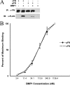
FIGURE 1.
FIGURE 1.
cFN and pFN bind BMP1. A, 80 ng of FLAG-tagged BMP1 was preincubated overnight with an equimolar amount of cFN or pFN. The complex was then immunoprecipitated (IP) with antisera raised against the N-terminal 70-kDa FN fragment (anti-70K), which recognizes both cFN and pFN. Immunoblotting (IB) was performed using either monoclonal anti-FLAG or polyclonal anti-70K antibody. B, BMP1 binding of cFN and pFN in ELISA. BMP1 at 3.4, 11.4, 34.1, 113.6, 340.6, and 1136.4 nm was incubated with cFN or pFN adsorbed to microtiter wells, as described under “Experimental Procedures.” The calculated KD values for BMP1 binding to cFN and pFN, as determined by Scatchard-type plots, were 110 ± 20 and 120 ± 30 nm, respectively. Values are expressed as the mean ± S.D. of three experiments.
FIGURE 2.
FIGURE 2.
BMP1 binds FN via its non-protease domain sequences. A, an immunoblot (IB) shows input amounts of cFN, FLAG-tagged versions of full-length BMP1, a form of BMP1 containing all domains except the protease domain (BMP1-D), and isolated BMP1 protease domain (BMP1-P). B, after overnight incubation of BMP1-D or BMP1-P with cFN, an immunoblot shows that the immunoprecipitation (IP) of cFN using anti-70K antibody pulls down full-length BMP1 and BMP1-D but not BMP1-P.
FIGURE 3.
FIGURE 3.
FN binds BMP1 at multiple sites. FLAG-tagged BMP1 was preincubated overnight with various truncated forms of FN followed by immunoprecipitation (IP). A, immunoblots (IB) show that immunoprecipitation of either the FN N-terminal 70-kDa fragment (70K) or the FN C-terminal fragment (III1-C) using anti-70K antibody or monoclonal LabMab anti-FN antibody, respectively, co-precipitated BMP1. B, immunoblots show that immunoprecipitation of III1-C(EDA+) or III1-C(EDA), with monoclonal LabMab antibody, co-precipitated similar amounts of BMP1. C, immunoblots show that immunoprecipitation of III1–14 with monoclonal LabMab antibody co-precipitated BMP1. D, a schematic is shown of the protein domain structure of FN and of truncated forms of FN used in this study. FN types I, II, and III repeats are shown. Alternatively spliced regions EDA, EDB, and V sequences are indicated, as are linker sequences (horizontal lines).
FIGURE 4.
FIGURE 4.
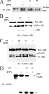
FN enhances BMP1 proteolytic processing of probiglycan, procollagen type I, and Chordin in vitro. Thirty-minute in vitro cleavage assays of probiglycan (A) or 6-h cleavage assays of type I procollagen (B and C) or Chordin (D) were performed in cFN-coated wells, with BSA-coated wells used as controls (A–C) or in the presence or absence of soluble cFN (D). A, probiglycan (ProBgn) and mature biglycan (Bgn) were detected by immunoblot (IB) using antibody (LF51) directed against sequences in the mature region of biglycan (20). B and C, procollagen, processing intermediates, and mature α1(I) chains were detected by immunoblot using C-telopeptide polyclonal antibody LF67 (20). Samples in B and C were run on 7.5 and 6.0% acrylamide SDS-PAGE gels, respectively, the latter to separate pC-α1(I) and pN-α1(I) processing intermediates. In B, in which reactions went somewhat further than in C, mature α1(I) chains are clearly discernable. D, BMP1 processing of C-terminal FLAG-tagged Chordin was monitored by immunoblot using anti-FLAG antibody. BMP1 cleaves Chordin at both N- and C-terminal sites (18). Bands for full-length Chordin (chd) and for a processing intermediate cleaved at the N-terminal but not the C-terminal site (C-chd) are marked but were not well separated in the 15% gel. Upon cleavage by BMP1, both bands disappeared, with the appearance of the 15-kDa Chordin C-terminal cleavage product (denoted by an asterisk), indicating complete Chordin processing.
FIGURE 5.
FIGURE 5.
Immunofluorescent co-localization of BMP1 and FN in fibroblast ECM. Paraformaldehyde-fixed cultures of human neonatal fibroblasts, either permeabilized or not permeabilized with Triton X-100, were immunostained with antibodies to FN (green) and BMP1 (red) and were stained with 4′,6-diamidino-2-phenylindole (DAPI, blue) for localization of nuclear DNA. The yellow signal indicates areas in which green and red signals overlap and, thus, areas of co-localization between FN and BMP1.
FIGURE 6.
FIGURE 6.
Detection of endogenous BMP1-FN complexes in cell culture. Conditioned medium from cultures of MG-63 cells pretreated 24 h with TGFβ1 was subjected to immunoprecipitation (IP) with anti-BMP1 antibody (lane labeled IP αBMP1 in panel A), anti-FN antibody (lane labeled IP αFN in panel B), or IgG (IP IgG-labeled lanes in both panels A and B) followed by immunoblotting (IB) with anti-FN or anti-BMP1 antibody. Input lanes represent 30 μl conditioned media not subjected to immunoprecipitation.
FIGURE 7.
FIGURE 7.
Reduced processing of endogenous BMP1 substrates in cultures of FN−/− MEFs. A, confluent FN+/− and FN−/− MEFs were incubated for 24 h in serum-free medium, and endogenous Chordin (chd) from the conditioned medium, concentrated by ethanol precipitation or with heparin-Sepharose, was probed via immunoblotting (IB) using anti-N-terminal-Chordin antibodies. Staining of ethanol-precipitated media samples with antibodies to secreted proteins sFRP2 and BMP1 shows similar levels of these two secreted proteins in FN+/− and FN−/− MEF media, arguing against overall differences in levels of secreted proteins despite different levels of detected Chordin. Staining of cell extracts with anti-α-tubulin antibody demonstrates that FN+/− and FN−/− MEF cultures contained similar numbers of cells despite different levels of detected Chordin from conditioned media. B, confluent FN+/− and FN−/− MEFs were treated for 24 h with 50 μg/ml ascorbate and then incubated for 24 h in serum-free medium containing 50 μg/ml ascorbate followed by harvesting of media and cell layer samples, which were then assayed via Western blot using LF67 antibody directed against the C-telopeptide of the α1(I) collagen chain. Positions are shown for procollagen, pNα1(I) chains ((pN), processing intermediates from which C-propeptides have been removed but which retain N-propeptides), pCα1(I) chains ((pC), processing intermediates from which N-propeptides have been removed but which retain C-propeptides), and mature, fully processed, α1(I) collagen chains. Reduced processing of the C-propeptides of type I procollagen and decreased deposition of mature collagen were observed in FN−/− MEF cultures compared with cultures of FN+/− MEFs, indicating dramatically reduced pCP activity for BMP1-like proteinases. C, confluent FN+/− and FN−/− MEFs were incubated for 24 h in serum-free medium, and probiglycan processing was examined in conditioned medium by Western blot using antibody LF51 directed against sequences within mature biglycan (20). Pro and mature forms of biglycan are denoted in the figure as ProBgn and Bgn, respectively. Probiglycan is less processed in FN−/− MEF cultures than in FN+/− MEF cultures.
FIGURE 8.
FIGURE 8.
Binding of endogenous FN and Chordin in cell culture. Conditioned medium from cultures of MG-63 cells pretreated 24 h with TGFβ1 was subjected to immunoprecipitation (IP) with anti-Chordin antibody (lane labeled chd in panel A), anti-FN antibody (lane labeled FN in panel B), or IgG (IgG-labeled lanes in panels A and B) followed by immunoblotting (IB) with anti-FN or anti-Chordin antibody. Input lanes represent 30 μl of conditioned media not subjected to immunoprecipitation.

Similar articles

Cited by

References

    1. Pankov R., Yamada K. M. (2002) J. Cell Sci. 115, 3861–3863 - PubMed
    1. Hynes R. O. (1990) Fibronectins, Springer-Verlag New York Inc., New York
    1. Magnusson M. K., Mosher D. F. (1998) Arterioscler. Thromb. Vasc. Biol. 18, 1363–1370 - PubMed
    1. Mao Y., Schwarzbauer J. E. (2005) Matrix Biol. 24, 389–399 - PubMed
    1. Ffrench-Constant C. (1995) Exp. Cell Res. 221, 261–271 - PubMed

Publication types

MeSH terms