. 2009 Sep 11;284(37):25077-86.
doi: 10.1074/jbc.M109.022061. Epub 2009 Jul 18.

Identification of linear epitopes in Bacillus anthracis protective antigen bound by neutralizing antibodies

Affiliations

Identification of linear epitopes in Bacillus anthracis protective antigen bound by neutralizing antibodies

Nareen Abboud et al. J Biol Chem. .

Abstract

Protective antigen (PA), the binding subunit of anthrax toxin, is the major component in the current anthrax vaccine, but the fine antigenic structure of PA is not well defined. To identify linear neutralizing epitopes of PA, 145 overlapping peptides covering the entire sequence of the protein were synthesized. Six monoclonal antibodies (mAbs) and antisera from mice specific for PA were tested for their reactivity to the peptides by enzyme-linked immunosorbent assays. Three major linear immunodominant B-cell epitopes were mapped to residues Leu(156) to Ser(170), Val(196) to Ile(210), and Ser(312) to Asn(326) of the PA protein. Two mAbs with toxin-neutralizing activity recognized two different epitopes in close proximity to the furin cleavage site in domain 1. The three-dimensional complex structure of PA and its neutralizing mAbs 7.5G and 19D9 were modeled using the molecular docking method providing models for the interacting epitope and paratope residues. For both mAbs, LeTx neutralization was associated with interference with furin cleavage, but they differed in effectiveness depending on whether they bound on the N- or C-terminal aspect of the cleaved products. The two peptides containing these epitopes that include amino acids Leu(156)-Ser(170) and Val(196)-Ile(210) were immunogenic and elicited neutralizing antibody responses to PA. These results identify the first linear neutralizing epitopes of PA and show that peptides containing epitope sequences can elicit neutralizing antibody responses, a finding that could be exploited for vaccine design.


Figures

FIGURE 1.
FIGURE 1.
Inverse antibody titers of BALB/c mice immunized with GalXM-PA conjugate as measured by ELISA. Two of the five female BALB/c mice (each bar represents one mouse) were initially immunized with 50 μl and one mouse was immunized with 100 μl of GalXM-PA conjugate in CFA on day 0. All mice were boosted with 50 μl of the conjugate in IFA at day 14. Sera were assayed for IgG anti-PA antibodies by ELISA. Inset, schematic of ELISA configuration used.
FIGURE 2.
FIGURE 2.
Western immunoblot analysis of mAbs binding to PA. The samples were separated by reduced SDS-10% polyacrylamide gels and then transferred onto nitrocellulose membrane. The membranes were then incubated with mAbs 29H, 16A12, 19D9, or 20G7 as the primary antibody and a horseradish peroxidase-conjugated goat isotype-specific antibody as the secondary antibody. The bands were visualized by ECL chemiluminescence kit. Lane 1, rPA undigested; lane 2, rPA + furin.
FIGURE 3.
FIGURE 3.
Analysis of cellular toxicity in the presence of anti-PA mAbs by MTT assay. mAb 19D9 protects J774 macrophage monolayers against LeTx-mediated toxicity. mAbs 2H9, 16A12, and 20G7 had no inhibitory effect against LeTx-mediated toxicity. MTT assays were done three times with similar results.
FIGURE 4.
FIGURE 4.
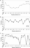
An alanine walk to determine the critical residues in three identified epitopes. Each residue extending from Leu156–Ser170 (A), Val196–Ile210 (B), or Ser312–Asn326 (C) was altered in turn to Ala or Gly (in the case if an Ala was present), and their reactivities with mAbs 7.5G, 29H, 19D9, and 20G7 were determined by ELISA. The absorbance readings of relative binding of mAbs to the alanine-substituted peptides is expressed as % binding of each altered peptide with respect to wild-type peptide, where the latter was considered the base-line maximum binding level. The background (Bkgd) of each mAb was determined in parallel, by using streptavidin-coated, peptide-free wells. The average value of three experiments is shown.
FIGURE 5.
FIGURE 5.
Cleavage of PA83 with furin in the absence and presence of mAb 19D9. The mAb 19D9 was incubated with PA for 1 h at room temperature prior to the adding of protease. The samples were separated by reduced SDS-10% polyacrylamide gels and stained with Coomassie Blue. MW, molecular weight marker; lane 1, PA undigested; lanes 2–5, PA + furin; lanes 6–9, PA + furin + mAb.
FIGURE 6.
FIGURE 6.
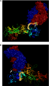
Molecular modeling analysis of mAbs 7.5G and 19D9. A, docking of 7.5G mAb on PA protein. B, docking of 19D9 mAb on PA protein. PA protein is depicted as a ribbon representation, whereas the epitopes, which are found in domain 1, are shown as yellow spheres. Arrows represent the furin site. mAbs are shown as space-filling models, where the red represents the heavy chain and the blue represents the light chain of the variable region.
FIGURE 7.
FIGURE 7.
Anti-peptide antibodies in MAP-D5- and MAP-E1-immunized mice. Female BALB/c mice (five animals per group) were immunized with 100 μg of MAP-D5 or MAP-E1 in CFA on day 0, and boosted with MAP-D5 or MAP-E1 in IFA on days 7 and 14, whereas control mice were immunized with MAP core in adjuvant. Sera from the different time points were diluted 1:500 and assayed for IgG anti-peptide antibodies by ELISA. Each point represents the average of five mice per treatment group. Inset, schematic of ELISA configuration used.
FIGURE 8.
FIGURE 8.
IgG subclasses of anti-MAP-D5 and anti-MAP-E1 antibodies induced following immunization with MAP-D5 and MAP-E1. The subclasses of the IgG anti-peptide antibodies in day +49 sera from five MAP-D5-immunized mice and five MAP-E1-immunized mice were measured by ELISA.
FIGURE 9.
FIGURE 9.
Anti-PA antibodies in MAP-D5- and MAP-E1-immunized mice and LeTx-neutralizing activity on J774 macrophages. A, sera from day +49 were assayed for IgG anti-PA antibodies by ELISA. The values represent geometric means ± S.D. from five mice per treatment group. B, day +49 serum from mice immunized with MAP-D5 and MAP-E1 confer moderate protection to J774 macrophage monolayers against LeTx-mediated toxicity. Bars represent arithmetic mean of the highest neutralization titers from five mice per treatment group. MTT assays were done three times with similar results. Anti-MAP core sera conferred no protection.

Similar articles

Cited by

References

    1. Barth H., Aktories K., Popoff M. R., Stiles B. G. (2004) Microbiol. Mol. Biol. Rev. 68, 373–402 - PMC - PubMed
    1. Inglesby T. V., O'Toole T., Henderson D. A., Bartlett J. G., Ascher M. S., Eitzen E., Friedlander A. M., Gerberding J., Hauer J., Hughes J., McDade J., Osterholm M. T., Parker G., Perl T. M., Russell P. K., Tonat K. (2002) J. Am. Med. Assoc. 287, 2236–2252 - PubMed
    1. Abboud N., Casadevall A. (2008) Clin. Vaccine Immunol. 15, 1115–1123 - PMC - PubMed
    1. Flick-Smith H. C., Walker N. J., Gibson P., Bullifent H., Hayward S., Miller J., Titball R. W., Williamson E. D. (2002) Infect. Immun. 70, 1653–1656 - PMC - PubMed
    1. Gubbins M. J., Berry J. D., Corbett C. R., Mogridge J., Yuan X. Y., Schmidt L., Nicolas B., Kabani A., Tsang R. S. (2006) FEMS Immunol. Med. Microbiol. 47, 436–443 - PMC - PubMed

Publication types

LinkOut - more resources