. 2009 Sep 18;284(38):25569-75.
doi: 10.1074/jbc.M109.019786. Epub 2009 Jul 18.

Adiponectin inhibits pro-inflammatory signaling in human macrophages independent of interleukin-10

Affiliations

Adiponectin inhibits pro-inflammatory signaling in human macrophages independent of interleukin-10

Eduardo J Folco et al. J Biol Chem. .

Abstract

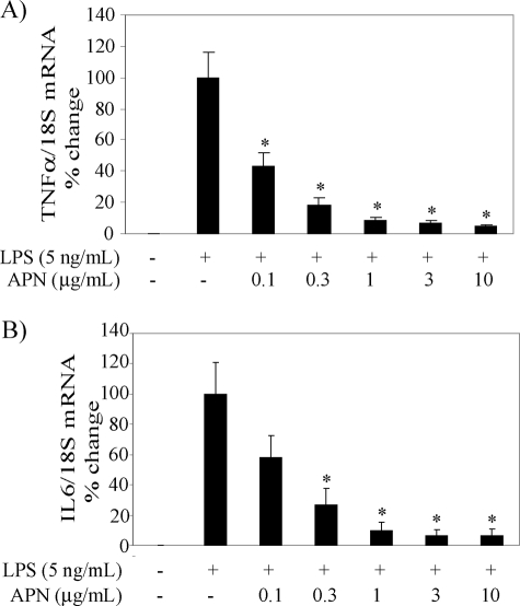
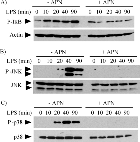
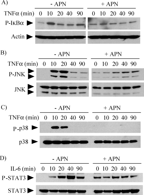
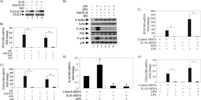
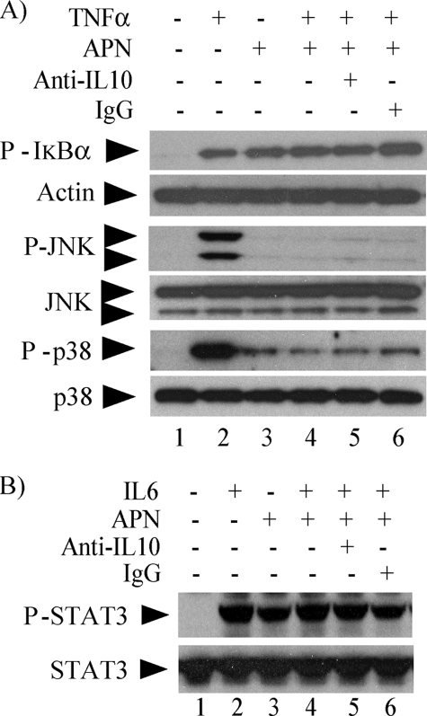
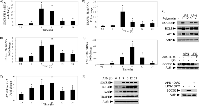
Macrophages participate pivotally in the pathogenesis of many chronic inflammatory diseases including atherosclerosis. Adiponectin, a vasculoprotective molecule with insulin-sensitizing and anti-atherogenic properties, suppresses pro-inflammatory gene expression in macrophages by mechanisms that remain incompletely understood. This study investigated the effects of adiponectin on major pro-inflammatory signaling pathways in human macrophages. We demonstrate that pretreatment of these cells with adiponectin inhibits phosphorylation of nuclear factor kappaB inhibitor (IkappaB), c-Jun N-terminal kinase (JNK), and p38 mitogen-activated protein kinase (MAPK), induced by either lipopolysaccharide (LPS) or tumor necrosis factor (TNF) alpha, as well as STAT3 phosphorylation induced by interleukin-6 (IL6). Antagonism of IL10 by either neutralizing antibodies or siRNA-mediated silencing did not abrogate the anti-inflammatory actions of adiponectin, indicating that the ability of adiponectin to render human macrophages tolerant to various pro-inflammatory stimuli does not require this cytokine. A systematic search for adiponectin-inducible genes with established anti-inflammatory properties revealed that adiponectin augmented the expression of A20, suppressor of cytokine signaling (SOCS) 3, B-cell CLL/lymphoma (BCL) 3, TNF receptor-associated factor (TRAF) 1, and TNFAIP3-interacting protein (TNIP) 3. These results suggest that adiponectin triggers a multifaceted response in human macrophages by inducing the expression of various anti-inflammatory proteins that act at different levels in concert to suppress macrophage activation.


Figures

FIGURE 1.
FIGURE 1.
Adiponectin attenuates TNFα and IL6 expression in human macrophages stimulated with LPS. Cells were treated with the indicated concentrations of adiponectin (APN) for 18 h and subsequently stimulated with 5 ng/ml LPS for 6 h. RT-qPCR measured mRNA levels for TNFα and IL6 as described under “Experimental Procedures.” Data are expressed relative to the values of treatment with LPS alone (100%) in means ± S.E. (n = 3). mRNA levels of 18S served as internal control for adjustment between samples. *, p < 0.05 versus LPS.
FIGURE 2.
FIGURE 2.
Adiponectin limits phosphorylation of IκB, JNK, and p38 in human macrophages stimulated with LPS. Cells were incubated without (left lanes) or with (right lanes) 10 μg/ml adiponectin for 18 h and subsequently stimulated with 5 ng/ml LPS for the indicated periods of time. Whole-cell lysates were fractionated by SDS-PAGE and immunoblotted with antibodies to phospho-IκB (A), phospho-JNK (B), or phospho-p38 (C). Actin, total JNK, and total p38 served as loading controls, respectively. Images are representative of three independent experiments on cells from different donors.
FIGURE 3.
FIGURE 3.
Adiponectin inhibits TNFα- and IL6-induced signaling in human macrophages. Cells were incubated without (left lanes) or with (right lanes) 10 μg/ml adiponectin for 18 h and subsequently stimulated with 50 ng/ml TNFα (A–C) or 5 ng/ml IL6 (D) for the indicated periods of time. Whole-cell lysates were fractionated by SDS-PAGE and immunoblotted with antibodies to phospho-IκB (A), phospho-JNK (B), phospho-p38 (C), or phospho-STAT3 (D). Actin, total JNK, total p38, and total STAT3 served as loading controls, respectively.
FIGURE 4.
FIGURE 4.
IL10 antagonism does not impair the anti-inflammatory actions of adiponectin in human macrophages stimulated with LPS. A, cells were incubated with no addition (lanes 1 and 2), 10 μg/ml anti-IL10 antibody (lane 3), or control IgG (lane 4) for 18 h and subsequently stimulated with (lanes 2–4) or without (lane 1) 10 ng/ml IL10 for 30 min. Whole-cell lysates were fractionated by SDS-PAGE and immunoblotted with antibodies to phospho-STAT3. Total STAT3 served as loading control. B and C, cells were treated with or without 10 μg/ml adiponectin for 18 h in the presence of anti-IL10 antibody or control IgG as indicated and subsequently stimulated with 5 ng/ml LPS for 6 h. RT-qPCR measured the levels of IP10 (B) and TNFα (C) mRNAs, using 18S as internal control for adjustment between samples. Data are expressed in means ± S.E. (n = 3) relative to the values of samples from cells without adiponectin or LPS treatment. *, p < 0.05. D, cells were incubated with (lanes 3–6) or without (lanes 1 and 2) 10 μg/ml adiponectin for 18 h in the presence of anti-IL10 antibody or control IgG as indicated and subsequently stimulated with 5 ng/ml LPS for 40 min. Whole-cell lysates were fractionated by SDS-PAGE and immunoblotted with antibodies to phospho-IκB (top), phospho-JNK (center), or phospho-p38 (bottom). Actin, total JNK, and total p38 served as loading controls, respectively. E, cells were transfected with IL10-specific or control siRNA as indicated. At 48 h after transfection, cells were incubated with or without 10 μg/ml adiponectin for 6 h. RT-qPCR measured IL10 mRNA levels using 18S as internal control for adjustment between samples. Data are expressed in means ± S.E. (n = 3) relative to the values of samples from cells transfected with control siRNA and left unstimulated. *, p < 0.05 versus control. F and G, cells were transfected with IL10-specific or control siRNA as indicated. At 48 h after transfection, cells were incubated with or without 10 μg/ml adiponectin for 18 h and subsequently stimulated with 5 ng/ml LPS for 6 h. RT-qPCR measured IP10 (F) and TNFα (G) mRNA levels using 18S as internal control for adjustment between samples. Data are expressed in means ± S.E. (n = 3) relative to the values of samples from cells transfected with control siRNA and left unstimulated.*, p < 0.05.
FIGURE 5.
FIGURE 5.
IL10 neutralization does not impair the actions of adiponectin in human macrophages stimulated with TNFα or IL6. A, cells were incubated with (lanes 3–6) or without (lanes 1 and 2) 10 μg/ml adiponectin for 18 h in the presence of anti-IL10 antibody (lane 5) or control IgG (lane 6) and subsequently stimulated with 50 ng/ml TNFα or left unstimulated for 10 min. Whole-cell lysates were fractionated by SDS-PAGE and immunoblotted with antibodies to phospho-IκB (top), phospho-JNK (middle), or phospho-p38 (bottom). Actin, total JNK, and total p38 served as loading controls, respectively. B, cells were incubated with (lanes 3–6) or without (lanes 1 and 2) 10 μg/ml adiponectin for 18 h in the presence of anti-IL10 antibody (lane 5) or control IgG (lane 6) and subsequently stimulated with 5 ng/ml IL6 or left unstimulated for 40 min. Whole-cell lysates were fractionated by SDS-PAGE and immunoblotted with antibodies to phospho-STAT3. Total STAT3 served as loading control.
FIGURE 6.
FIGURE 6.
Adiponectin induces expression of various anti-inflammatory molecules in human macrophages. Human macrophages were incubated with 10 μg/ml adiponectin for the indicated periods of time. In A–E, RT-qPCR measured the levels of the indicated mRNAs, using 18S as internal control for adjustment between samples. Data are expressed in means ± S.E. (n = 3) relative to the values of samples from vehicle-treated cells at the respective time points. *, p < 0.05. In F, whole-cell lysates were fractionated by SDS-PAGE and immunoblotted with the indicated antibodies, using actin as loading control. In G, cells were pretreated with 1 μg/ml polymyxin (top) or 25 μg/ml of either anti-TLR4 antibodies or control IgG (middle), followed by stimulation with 5 ng/ml LPS or 10 μg/ml adiponectin for 3 h. In G (bottom), adiponectin or LPS were subjected to a 20-min pretreatment at 100 °C before addition to the cells and incubation for 3 h. Whole-cell lysates were fractionated by SDS-PAGE and immunoblotted with the indicated antibodies, using actin as loading control.

References

    1. Rocha V. Z., Libby P. (2009) Nat. Rev. Cardiol. 6, 399–409 - PubMed
    1. Scherer P. E. (2006) Diabetes 55, 1537–1545 - PubMed
    1. Goldstein B. J., Scalia R. G., Ma X. L. (2009) Nat. Clin. Pract. Cardiovasc. Med. 6, 27–35 - PMC - PubMed
    1. Okamoto Y., Kihara S., Funahashi T., Matsuzawa Y., Libby P. (2006) Clin. Sci. 110, 267–278 - PubMed
    1. Zhu W., Cheng K. K., Vanhoutte P. M., Lam K. S., Xu A. (2008) Clin. Sci. 114, 361–374 - PubMed

Publication types

MeSH terms

Substances