. 2009 Sep 11;284(37):25135-48.
doi: 10.1074/jbc.M109.013292. Epub 2009 Jul 18.

Dioxin receptor deficiency impairs angiogenesis by a mechanism involving VEGF-A depletion in the endothelium and transforming growth factor-beta overexpression in the stroma

Affiliations

Dioxin receptor deficiency impairs angiogenesis by a mechanism involving VEGF-A depletion in the endothelium and transforming growth factor-beta overexpression in the stroma

Angel Carlos Roman et al. J Biol Chem. .

Abstract

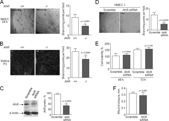
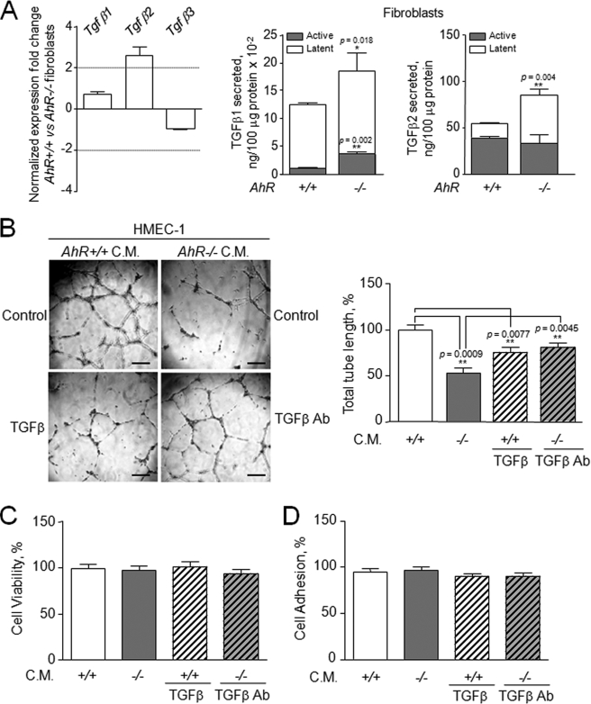
Angiogenesis has key roles in development and in the progression of human diseases such as cancer. Consequently, identifying the novel markers and regulators of angiogenesis is a critical task. The dioxin receptor (AhR) contributes to vascular homeostasis and to the endothelial response to toxins, although the mechanisms involved are largely uncharacterized. Here, we show that AhR-null mice (AhR(-/-)) have impaired angiogenesis in vivo that compromises tumor xenograft growth. Aortic rings emigration experiments and RNA interference indicated that AhR(-/-) endothelial cells failed to branch and to form tube-like structures. Such a phenotype was found to be vascular endothelial growth factor (VEGF)-dependent, as AhR(-/-) aortic endothelial cells (MAECs) secreted lower amounts of active VEGF-A and their treatment with VEGF-A rescued angiogenesis in culture and in vivo. Further, the addition of anti-VEGF antibody to AhR(+/+) MAECs reduced angiogenesis. Treatment under hypoxic conditions with 2-methoxyestradiol suggested that HIF-1alpha modulates endothelial VEGF expression in an AhR-dependent manner. Importantly, AhR-null stromal myofibroblasts produced increased transforming growth factor-beta (TGFbeta) activity, which inhibited angiogenesis in human endothelial cells (HMECs) and AhR(-/-) mice, whereas the co-culture of HMECs with AhR(-/-) myofibroblasts or with their conditioned medium inhibited branching, which was restored by an anti-TGFbeta antibody. Moreover, VEGF and TGFbeta activities cooperated in modulating angiogenesis, as the addition of TGFbeta to AhR(-/-) MAECs further reduced their low basal VEGF-A activity. Thus, AhR modulates angiogenesis through a mechanism requiring VEGF activation in the endothelium and TGFbeta inactivation in the stroma. These data highlight the role of AhR in cardiovascular homeostasis and suggest that this receptor can be a novel regulator of angiogenesis during tumor development.


Figures

FIGURE 1.
FIGURE 1.
AhR−/− mice have impaired tumor growth and inefficient angiogenesis. A, B16F10 mouse melanoma cells were injected subcutaneously in the dorsal area of AhR+/+ and AhR−/− mice, and tumors were collected at 7 and 14 days after grafting. Tumor volume was calculated as length × width2 × 0.4. B, tumors were fixed, sectioned, and stained with hematoxylin and eosin or used to detect blood vessels by immunofluorescence with an antibody for the endothelial marker CD31. Vessels were counted at each time point, and the results corresponding to tumors at 14 days were represented. Note that hematoxylin and eosin images appear dark because of the high levels of melanin expressed by B16F10 melanoma cells. C, Matrigel was prepared and injected subcutaneously in the dorsal area of AhR+/+ and AhR−/− mice. After 7 days, the Matrigel plugs formed were removed, washed, and photographed, and the angiogenesis recruited in each genotype was quantitated by measuring the content in hemoglobin. Hemoglobin content is represented as mg/ml in 100 mg of Matrigel. Light microscopy was done at room temperature on a Nikon E-400 microscope equipped with a Nikon L16 camera. A ×10 objective (0.25 numeric aperture) was used on Eukitt-mounted sections. Immunofluorescence was done at room temperature on a Nikon TE2000U microscope equipped with a Nikon DS-5 M digital camera. A ×10 objective (0.25 numeric aperture) was used on Mowiol-mounted sections. Data are shown as means ± S.D. Tumors were induced in both sides of five AhR+/+ and AhR−/− mice at each time point. Matrigel plugs were done in duplicate in at least four animals of each genotype.
FIGURE 2.
FIGURE 2.
AhR−/− mouse endothelial cells have a reduced potential for tubulogenesis and branching in aortic ring explants. A, schematic representation of the method used to calculate TTL and branching points in aortic ring explants and Matrigel plugs. Branching points are indicated by arrows. B, rings were prepared from the aortas of AhR+/+ and AhR−/− mice and placed in Matrigel plugs. After 7 days in culture, tube formation was determined by measuring TTL for the major sprouting vessels as described (47). Maximum outgrowth was also determined as the total distance emigrated by the mouse endothelial cells (MAEC) from the aortic ring (ar). C, tube formation was also analyzed by staining the aortic rings with 4,6-diamidino-2-phenylindole. Tube formation was calculated as the ratio between tube-oriented/dispersed endothelial cells. Light microscopy was done at room temperature on a Nikon E-400 microscope equipped with a Nikon L16 camera. Objectives used were: ×4 (0.10 numeric aperture) (A) and ×10 (0.25 numeric aperture) (B and C). Data are shown as means ± S.D. At least 15 aortic rings were analyzed from different mice of each genotype.
FIGURE 3.
FIGURE 3.
Down-modulation of AhR expression in mouse and human aortic endothelial cells reduces branching and tube formation. A, primary MAEC cultures were obtained by isolating endothelial cells emigrating from the aortic rings. Cells were plated in Matrigel, and branching was quantitated 24 h later. B, retinas were isolated from AhR+/+ and AhR−/− 3-day-old newborn mice and stained with isolectin B4-FITC to label blood vessels. Branching points were determined and plotted for each genotype. Measurements were taken near to the optic nerve, at the center of the retina. C, HMEC-1 cells were transfected by nucleoporation with an AhR siRNA or unspecific scramble RNA, and AhR protein levels were determined by Western immunoblotting 72 h later. The expression of β-actin was used as negative control for RNAi and as loading control for Western blotting. D, siRNA- or scramble RNA-transfected HMEC-1 cells were plated in Matrigel. Tube formation and branching were analyzed after 24 h as described under ”Experimental Procedures.“ E, cell viability was determined in HMEC-1 cells at 48 or 72 h after transfection with either scramble or AhR siRNA. Cells were stained with crystal violet and counted. F, HMEC-1 cells were transfected with scramble or AhR siRNA and grown to confluence. Serum-free culture medium was added, and wound healing performed as indicated under ”Experimental Procedures.“ Data represent the extent of wound closure under both experimental conditions. Light microscopy was done at room temperature on a Nikon E-400 microscope equipped with a Nikon L16 camera. Objectives used were: ×10 (0.25 numeric aperture) (A and D) and ×20 (0.40 numeric aperture) (B). Data are shown as means ± S.D. Branching was analyzed in at least five primary MAEC cultures obtained from aortic rings isolated from different mice. Retinas were isolated from eyes of three AhR+/+ and AhR−/− mice. HMEC-1 cells were transfected in triplicate with AhR siRNA or unspecific scramble RNA. Calibration bar corresponds to 100 μm.
FIGURE 4.
FIGURE 4.
AhR−/− MAECs have reduced expression and activity of the proangiogenesis factor, VEGF. A, total RNA was purified from AhR+/+ and AhR−/− MAECs and used to analyze Hif-1α, Vegf-A, Vegf-B, Plgf, Vegfr-1, and Vegfr-2 expression by real-time RT-PCR. Gene expression was normalized by Gapdh expression. Fold change represents the difference between AhR−/− MAECs and AhR+/+ MAECs. The oligonucleotide sequences used to amplify each gene are indicated in Table 1. Determinations were done in triplicate in at least three MAEC cultures. B, mRNA expression for the Vegf120, Vegf164, and Vegf188 isoforms was determined by real-time RT-PCR. Differences in expression between AhR−/− and AhR+/+ MAECs are indicated as -fold change. The oligonucleotide sequences used to amplify each gene are indicated in Table 1. C, the amount of VEGF-A activity secreted by AhR+/+ and AhR−/− MAEC and fibroblast cells was determined by ELISA in conditioned medium from each cell type and genotype. D, hypoxia was mimicked in AhR+/+ and AhR−/− MAEC cultures by treatment with 100 μm CoCl2 for 16 or 24 h, and Vegf expression was determined at the mRNA level as indicated under ”Experimental Procedures.“ Data were normalized and expressed as -fold change with respect to solvent (DMSO)-treated cultures. In some experiments, MAECs were co-treated with 100 μm CoCl2 and 5 μm 2-Me E2 for 24 h to induce HIF-1α degradation under hypoxic conditions. Vegf expression was normalized using β-actin instead of Gapdh to avoid potential effects of CoCl2 on the expression of the latter gene. The experiments were done in triplicate in three MAEC cultures. Data are shown as means ± S.D.
FIGURE 5.
FIGURE 5.
AhR down-modulation by siRNA decreases Hif-1α and Vegf-A expression in HMEC-1 cells. A, HMEC-1 cells were transfected with scramble of AhR-specific siRNA by nucleoporation. Total RNA was purified and analyzed for Hif-1α and Vegf-A mRNA expression by real-time RT-PCR using the oligonucleotides indicated in Table 1. Data were normalized, and fold change represents the difference between scramble- and AhR siRNA-transfected cultures. B, HMEC-1 cells were transfected as indicated in A, and some cultures were treated for 24 h with 5 μm 2-Me E2. Total RNA was purified and analyzed for Vegf165 mRNA expression as indicated in the legend for Fig. 4. The experiments were done in two different transfections using triplicate samples. Data are shown as means ± S.D.
FIGURE 6.
FIGURE 6.
VEGF activity modulates angiogenesis in an AhR-dependent manner in vivo and in culture. A, aortic rings (ar) were obtained from AhR+/+ and AhR−/− mice and plated in Matrigel plugs for 7 days. Sprouting of MAECs was analyzed by measuring the TTL as described (47). In some experiments, AhR+/+ or AhR−/− aortic rings were treated with anti-VEGF-A antibody (200 ng/ml) or recombinant VEGF-A164 protein (100 ng/ml), respectively. Light microscopy was done at room temperature on a Nikon E-400 microscope equipped with a Nikon L16 camera with a ×10 objective (0.25 numeric aperture). The experiments were performed in at least 10 aortic rings obtained from different AhR+/+ and AhR−/− mice. Magnified images are shown below the main panels. B, Matrigel plugs were injected in the dorsal skin of AhR+/+ or AhR−/− mice and left untreated or treated with anti-VEGF-A antibody (200 ng/ml) or recombinant VEGF-A164 protein (300 ng/ml), respectively. Blood vessel recruitment to Matrigel plugs was quantitated by measuring their hemoglobin content. Hemoglobin content is represented as mg/ml in 100 mg of Matrigel. At least eight aortic rings from different mice were used for each genotype and experimental condition. Four mice of each genotype were used for the in vivo Matrigel experiments. One flank of each mouse was left untreated as a control, and the other flank was treated as indicated. Data are shown as means ± S.D.
FIGURE 7.
FIGURE 7.
Co-culture of human endothelial HMEC-1 cells with AhR−/− fibroblasts inhibits tube formation. (A, scheme showing the experimental system used to co-culture HMEC-1 cells with fibroblasts. B, HMEC-1 cells grown in Matrigel plugs were co-cultured with AhR+/+ or AhR−/− fibroblasts, and their ability to form tubes was determined by measuring TTL as described (47). C, HMEC-1 cells were cultured in Matrigel plugs in the presence of medium conditioned (C.M.) by AhR+/+ or AhR−/− fibroblasts, and TTL was determined as above. Light microscopy was done at room temperature on a Nikon E-400 microscope equipped with a Nikon L16 camera with a ×10 objective (0.25 numeric aperture). The experiments were performed in four HMEC-1 cultures for each experimental condition. Data are shown as means ± S.D.
FIGURE 8.
FIGURE 8.
The addition of recombinant TGFβ or neutralizing anti-TGFβ antibody to medium conditioned by AhR−/− fibroblasts modulates angiogenesis of HMEC-1 cells. A, expression of Tgfβ-1, Tgfβ-2, and Tgfβ-3 isoforms was analyzed in AhR+/+ and AhR−/− fibroblasts by real-time RT-PCR using the oligonucleotides indicated in Table 1. Secretion of latent and active TGFβ-1 and TGFβ-2 proteins by AhR+/+ and AhR−/− fibroblasts to the culture medium was determined by specific ELISA as indicated under ”Experimental Procedures.“ To activate latent TGFβ, conditioned medium was treated for 10 min at room temperature with 165 mm HCl and then neutralized with NaOH. B, HMEC-1 were plated in Matrigel and treated with CM from AhR+/+ or AhR−/− fibroblasts, with AhR+/+ CM plus 10 ng/ml recombinant TGFβ, or with AhR−/− CM plus 1 μg/ml neutralizing anti-TGFβ antibody (Ab). TTL was determined as indicated (47). Light microscopy was done at room temperature on a Nikon E-400 microscope equipped with a Nikon L16 camera with a ×10 objective (0.25 numeric aperture). The experiments were performed in three HMEC-1 cultures for each experimental condition. Data are shown as means ± S.D. C, viability of HMEC-1 cells was determined under the same experimental conditions as those used in B. D, cell adhesion was quantitated in HMEC-1 cells under the experimental conditions used in B. Determinations were done in triplicate in at least two different HMEC-1 cultures. Data are shown as means ± S.D.
FIGURE 9.
FIGURE 9.
Medium conditioned by AhR−/− fibroblasts inhibits invasion of HMEC-1 cells, and modulation of VEGF and TGFβ levels affects blood vessel recruitment in vivo. A, HMEC-1 cells plated in Matrigel-coated Transwells were treated with CM from AhR+/+ or AhR−/− fibroblasts, with AhR+/+ CM plus 10 ng/ml recombinant TGFβ, or with AhR−/− CM plus 1 μg/ml neutralizing anti-TGFβ antibody (Ab). HMEC-1 invasion was quantitated by confocal microscopy taking measurements each 5 μm. Confocal microscopy was done at room temperature on a Zeiss LSM 510 apparatus. At least three Transwells were used for each genotype and experimental condition. B, Matrigel plugs were injected into the dorsal skin of AhR+/+ or AhR−/− mice and left untreated or treated with recombinant TGFβ (10 ng/ml) (AhR+/+ mice), neutralizing anti-TGFβ antibody (1 μg/ml) (AhR−/− mice), recombinant TGFβ (10 ng/ml) plus anti-VEGF-A antibody (200 ng/ml) (AhR+/+ mice), or neutralizing anti-TGFβ antibody (1 μg/ml) plus recombinant VEGF-A164 protein (300 ng/ml) (AhR−/− mice). After 7 days, the plugs were recovered, photographed, and used to quantify vessel formation by measuring their hemoglobin content. Hemoglobin content is represented as mg/ml in 100 mg of Matrigel. The experiments were repeated in at least four mice of each genotype. One flank of each mouse was used as an untreated control, and the other flank was treated as indicated. C, AhR+/+ and AhR−/− MAECs were left untreated or treated with neutralizing anti-TGFβ antibody (1 μg/ml) or recombinant TGFβ (10 ng/ml), respectively. VEGF-A activity secreted by these cells was measured by ELISA as indicated in the legend for Fig. 4. Data are shown as means ± S.D.

Similar articles

Cited by

References

    1. Lloyd R. V., Vidal S., Horvath E., Kovacs K., Scheithauer B. (2003) Microsc. Res. Tech. 60, 244–250 - PubMed
    1. Ferrara N., Alitalo K. (1999) Nat. Med. 5, 1359–1364 - PubMed
    1. Gerhardt H., Golding M., Fruttiger M., Ruhrberg C., Lundkvist A., Abramsson A., Jeltsch M., Mitchell C., Alitalo K., Shima D., Betsholtz C. (2003) J. Cell Biol. 161, 1163–1177 - PMC - PubMed
    1. Tammela T., Zarkada G., Wallgard E., Murtomäki A., Suchting S., Wirzenius M., Waltari M., Hellström M., Schomber T., Peltonen R., Freitas C., Duarte A., Isoniemi H., Laakkonen P., Christofori G., Ylä-Herttuala S., Shibuya M., Pytowski B., Eichmann A., Betsholtz C., Alitalo K. (2008) Nature 454, 656–660 - PubMed
    1. Childs S., Chen J. N., Garrity D. M., Fishman M. C. (2002) Development 129, 973–982 - PubMed

Publication types

LinkOut - more resources