. 2009 Oct 23;284(43):29784-97.
doi: 10.1074/jbc.M109.038497. Epub 2009 Jul 18.

Structural basis for T cell alloreactivity among three HLA-B14 and HLA-B27 antigens

Affiliations

Structural basis for T cell alloreactivity among three HLA-B14 and HLA-B27 antigens

Pravin Kumar et al. J Biol Chem. .

Abstract

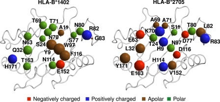
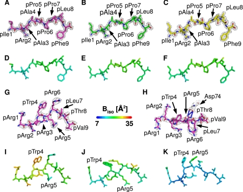
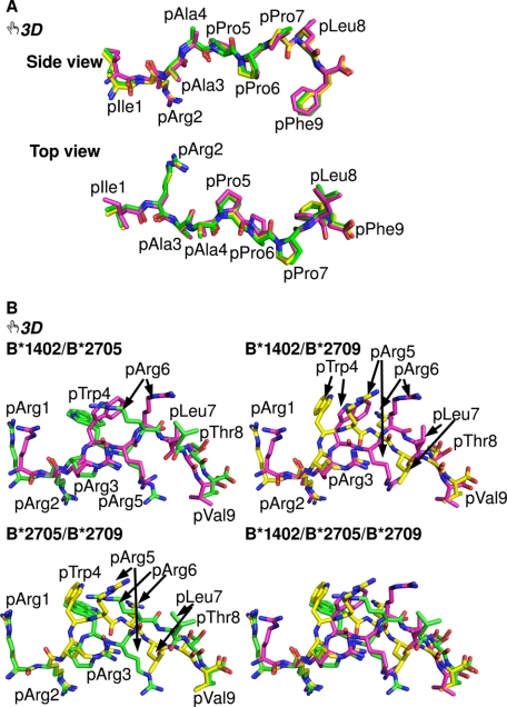
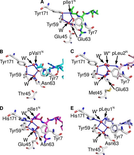
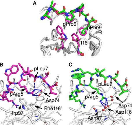
The existence of cytotoxic T cells (CTL) cross-reacting with the human major histocompatibility antigens HLA-B14 and HLA-B27 suggests that their alloreactivity could be due to presentation of shared peptides in similar binding modes by these molecules. We therefore determined the crystal structures of the subtypes HLA-B*1402, HLA-B*2705, and HLA-B*2709 in complex with a proven self-ligand, pCatA (peptide with the sequence IRAAPPPLF derived from cathepsin A (residues 2-10)), and of HLA-B*1402 in complex with a viral peptide, pLMP2 (RRRWRRLTV, derived from latent membrane protein 2 (residues 236-244) of Epstein-Barr virus). Despite the exchange of 18 residues within the binding grooves of HLA-B*1402 and HLA-B*2705 or HLA-B*2709, the pCatA peptide is presented in nearly identical conformations. However, pLMP2 is displayed by HLA-B*1402 in a conformation distinct from those previously found in the two HLA-B27 subtypes. In addition, the complexes of HLA-B*1402 with the two peptides reveal a nonstandard, tetragonal mode of the peptide N terminus anchoring in the binding groove because of the exchange of the common Tyr-171 by His-171 of the HLA-B*1402 heavy chain. This exchange appears also responsible for reduced stability of HLA-B14-peptide complexes in vivo and slow assembly in vitro. The studies with the pCatA peptide uncover that CTL cross-reactive between HLA-B14 and HLA-B27 might primarily recognize the common structural features of the bound peptide, thus neglecting amino acid replacements within the rim of the binding grooves. In contrast, structural alterations between the three complexes with the pLMP2 peptide indicate how heavy chain polymorphisms can influence peptide display and prevent CTL cross-reactivity between HLA-B14 and HLA-B27 antigens.


Figures

FIGURE 1.
FIGURE 1.
Amino acid sequence differences among B*1402 and B*2705 HC. The 18 residues distinguishing the two subtypes are all located in or in the immediate vicinity of the peptide-binding groove. B*2705 differs from B*2709 only by a D116H exchange (not shown). The residues are indicated by spheres with volumes roughly proportional to the volumes of the respective amino acid side chain in solution (77). The spheres are colored according to the biochemical properties of the respective amino acids, as indicated at the bottom of the image.
FIGURE 2.
FIGURE 2.
General properties of the pCatA and pLMP2 peptides bound to three HLA-B subtypes. A–C, final 2Fo − Fc electron density of pCatA conformations in B*1402 (A), B*2705 (B), and B*2709 (C), contoured at 1.5σ level. d–F, pCatA bound by B*1402 (D), B*2705 (E), and B*2709 (F) color-coded by isotropic B-factor. G and H, final 2Fo − Fc electron density of pLMP2 in B*1402 shown from the α2-helix (G) or the top of the binding groove (H), contoured at 1.5σ level. Electron density is depicted only for one molecule in the asymmetric unit. I–K, pLMP2 bound by B*1402 (I), B*2705 (J), and B*2709 (K) color-coded by isotropic B-factor. In all figures, unless mentioned otherwise, peptides bound to B*1402 are shown in violet, and peptides bound to B*2705 are shown in green, and peptides bound to B*2709 in are shown in yellow.
FIGURE 3.
FIGURE 3.
Interactive three-dimensional comparison of the pCatA and pLMP2 peptides bound to three HLA-B molecules. An overlay of the conformations of pCatA in B*1402 (violet), B*2705 (green), and B*2709 (yellow) is provided in A and for pLMP2 in B. By clicking on each of the two-dimensional images in the PDF version of the article, the three-dimensional functions for a given peptide become available (they can be terminated by right-clicking on the three-dimensional display and choosing the “Disable three-dimensional” function). In each case, the model tree icon allows the view of individual components (with their designations shown on the left) of the three-dimensional model, and offers a preselected “tour” of the model. During the tour (with 6 and 10 images for B*1402 or B*2705, respectively), each of the models can be manipulated individually using the mouse (the tools to rotate, pan, or zoom an image can be selected through the toolbar or the contextual menu). A better understanding of many structural characteristics of the models can be obtained by “playing” with the structures. For example, the different contacts made by pArg5 in the binding grooves of B*1402 and B*2705, respectively, are easily compared with each other, as Asp-74 and the polymorphic residue 116 (Phe in B*1402, Asp in B*2705, and His in B*2709) are shown, along with residue 97 (views 6–10). Colors of the peptides as in Fig. 2.
FIGURE 4.
FIGURE 4.
HLA-B subtype-dependent anchoring of peptide N termini. The crucial role of the Y171H exchange for the anchoring of peptide N termini is revealed by comparing the A pockets of B*2705·pCatA (A), B*3501 bound to the octameric nef peptide (B), A*0201 complexed with an N-terminally truncated peptide (C), B*1402·pCatA (D), and B*5101·KM1 (E). Whereas the first three structures (all with Tyr-171) show a pentagonal hydrogen bonding network, the subtypes B*1402 and B*5101 anchor the peptide N terminus only indirectly, via a water molecule (W′) which is also contacted by His-171NE, resulting in the formation of a tetragonal network of H-bonds. HC residues are colored gray, and peptide residues are in different colors.
FIGURE 5.
FIGURE 5.
Molecular surfaces of the pCatA and pLMP2 peptides bound to three HLA-B subtypes. Shown are molecular surface representations of B*1402 (A and D), B*2705 (B and E), and B*2709 (C and F) complexed with the pCatA peptide (A–C) or the pLMP2 peptide (D–F), respectively, as viewed by an approaching TCR. Colors of the peptides are as in Fig. 2. In each image, electrostatic surfaces are shown only for the respective HC, with red indicating a negative and blue a positive charge. Gray areas are uncharged. A number of crucial peptide or HC residues (see text) are indicated.
FIGURE 6.
FIGURE 6.
Differential anchoring of the middle of the pCatA and pLMP2 peptides. The bulky residues at HC positions 97 (Trp) and 116 (Phe) in the B*1402 subtype lead to differential contacts when compared with B*2705 (Asn-97 and Asp-116). The structures with pCatA (violet) (A) and pLMP2 (B) (green, A and C) are shown. Whereas pPro6 of pCatA is shifted only slightly into the groove in case of B*1402, the smaller side chains of residues 97 and 116 do not contact pPro6. The pArg5 residue of the pLMP2 peptide forms a bidentate salt bridge with Asp-74 and a hydrogen bond with pLeu7 in B*1402, whereas salt bridges with Asp-116 characterize the noncanonical pLMP2 conformation in case of B*2705.

Similar articles

Cited by

References

    1. Lindahl K. F., Wilson D. B. (1977) J. Exp. Med. 145, 508–522 - PMC - PubMed
    1. Suchin E. J., Langmuir P. B., Palmer E., Sayegh M. H., Wells A. D., Turka L. A. (2001) J. Immunol. 166, 973–981 - PubMed
    1. Afzali B., Lechler R. I., Hernandez-Fuentes M. P. (2007) Tissue Antigens 69, 545–556 - PubMed
    1. Kamoun M., Holmes J. H., Israni A. K., Kearns J. D., Teal V., Yang W. P., Rosas S. E., Joffe M. M., Li H., Feldman H. I. (2008) Proc. Natl. Acad. Sci. U.S.A. 105, 18883–18888 - PMC - PubMed
    1. Morris G. P., Allen P. M. (2009) J. Immunol. 182, 6639–6643 - PMC - PubMed

Publication types