

Review
doi: 10.1038/nrn2683.
The genetic and molecular regulation of sleep: from fruit flies to humans
Affiliations
- PMID: 19617891
- PMCID: PMC2767184
- DOI: 10.1038/nrn2683
Item in Clipboard
Review
The genetic and molecular regulation of sleep: from fruit flies to humans
Nat Rev Neurosci.
2009 Aug.
Abstract
It has been known for a long time that genetic factors affect sleep quantity and quality. Genetic screens have identified several mutations that affect sleep across species, pointing to an evolutionary conserved regulation of sleep. Moreover, it has also been recognized that sleep affects gene expression. These findings have given valuable insights into the molecular underpinnings of sleep regulation and function that might lead the way to more efficient treatments for sleep disorders.
Figures
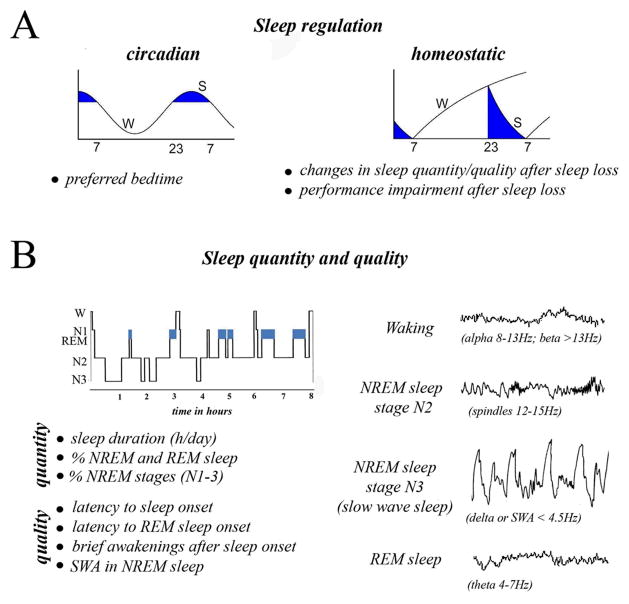
Sleep phenotypes. Sleep phenotypes such as regulation, sleep duration (quantity), or sleep intensity (quality) may reflect different aspects of sleep. A) Sleep regulation. Blue areas indicate time of day most conducive to sleep in humans, due to the combined effect of the circadian system, which consolidates sleep during the dark phase, and the homeostatic system, which increases sleep pressure as a function of waking duration. B) Sleep quantity and quality. Top left, night distribution of sleep stages in adult humans. Right, representative EEG traces in waking, NREM sleep and REM sleep. The waking “activated” EEG is dominated by low voltage fast activity in the beta (>13Hz) and alpha (8–13Hz) range. NREM sleep comprises a transitional stage 1 (N1, not shown), when alpha activity disappears, followed by stage 2 (N2), rich in sleep spindles, and then N3 or slow wave sleep (SWS, also called stages 3+4), when the EEG shows prominent slow waves . The higher the number of slow waves, the deeper is NREM sleep, i.e. the more difficult is to wake up. Sleep spindles are waxing and waning oscillations of thalamic origin whose frequency (12–15 Hz) is comprised within the sigma band (12–16Hz), while slow waves are of cortical origin and are comprised within the delta band, also called slow wave activity (SWA, <4.5Hz).
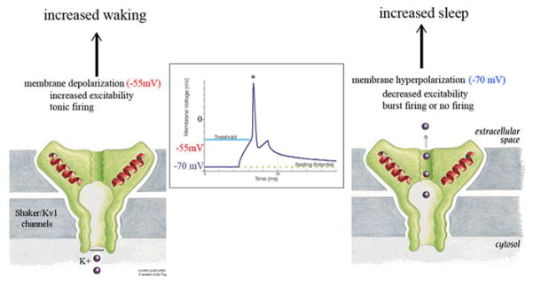
Proposed mechanism for the short sleeping phenotype caused by loss of function mutations of Shaker/Kv1 potassium channels. Normally, the opening of these channels allow K+ ions to exit the neuron, bringing the membrane potential to more hyperpolarized (more negative) levels, close to the resting membrane potential. Mutations that reduce the total number of these channels, and/or decrease the time the channel can remain open, tend to bring the membrane potential to more positive (depolarized levels), closer to the threshold for firing an action potential (* in the figure).
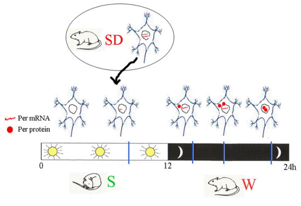
Model showing changes in the expression of Period genes (mRNA and protein) as a function of the 24-hour cycle and in response to sleep deprivation (SD): mRNA levels grow during the day, while protein levels peak at night, when Per enters the nucleus and blocks its own expression. These circadian changes are similar regardless of whether the animal is diurnal (like flies, which sleep mainly at night) or nocturnal (like rats, which sleep mostly during the day). Rodents forced to stay awake during the day show increased Per mRNA levels in the cerebral cortex. In both flies and mammals, however, sleep deprivation usually does not reset the circadian clock, i.e. does not cause phase shifts.
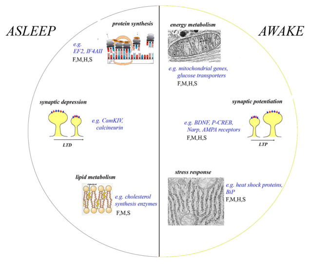
Schematic representation of the major functional categories of genes whose expression is higher in the rat brain after several hours of wakefulness (including after 3–8 hours of sleep deprivation) or after several hours of sleep ,,. F, M, H, S indicate when changes in the same functional category are also present in the brain of fruit flies, mice, Djungarian hamsters (Tom DeBoer, Irene Tobler, Chiara Cirelli, unpublished results) and sparrows, respectively.
References
-
- Tobler I, et al. Altered circadian activity rhythms and sleep in mice devoid of prion protein. Nature. 1996;380:639–642. First study in mice to show that a null mutation affects sleep regulation. - PubMed
-
- Lin L, et al. The sleep disorder canine narcolepsy is caused by a mutation in the hypocretin (orexin) receptor 2 gene. Cell. 1999;98:365–376. Seminal study that identifies the autosomal recessive mutation responsible for canine narcolepsy. - PubMed
-
- Chemelli RM, et al. Narcolepsy in orexin knockout mice: Molecular genetics of sleep regulation. Cell. 1999;98:437–451. This study shows that mice lacking hypocretin/orexin have a narcolepsy-like phenotype. - PubMed
Publication types
MeSH terms
Substances
Grants and funding
LinkOut - more resources
Full Text Sources
Molecular Biology Databases
