. 2009 Jul 20;4(7):e6314.
doi: 10.1371/journal.pone.0006314.

Remission of invasive, cancer stem-like glioblastoma xenografts using lentiviral vector-mediated suicide gene therapy

Affiliations

Remission of invasive, cancer stem-like glioblastoma xenografts using lentiviral vector-mediated suicide gene therapy

Peter C Huszthy et al. PLoS One. .

Abstract

Background: Glioblastoma is the most frequent and most malignant primary brain tumor with a poor prognosis. The translation of therapeutic strategies for glioblastoma from the experimental phase into the clinic has been limited by insufficient animal models, which lack important features of human tumors. Lentiviral gene therapy is an attractive therapeutic option for human glioblastoma, which we validated in a clinically relevant animal model.

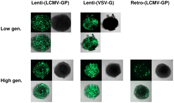
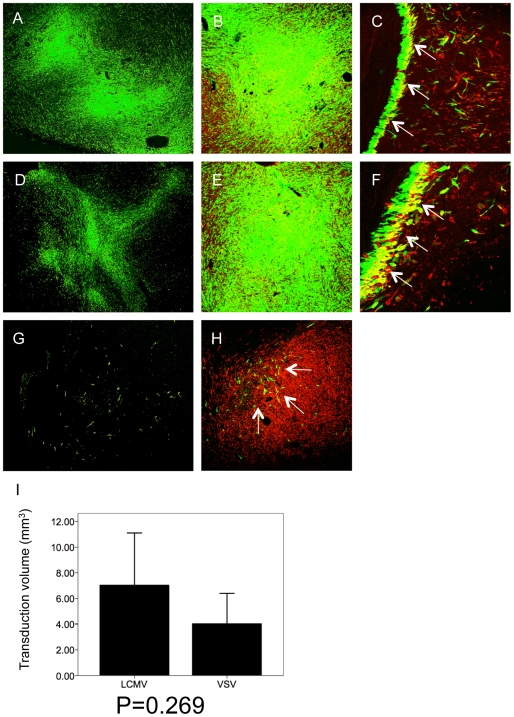
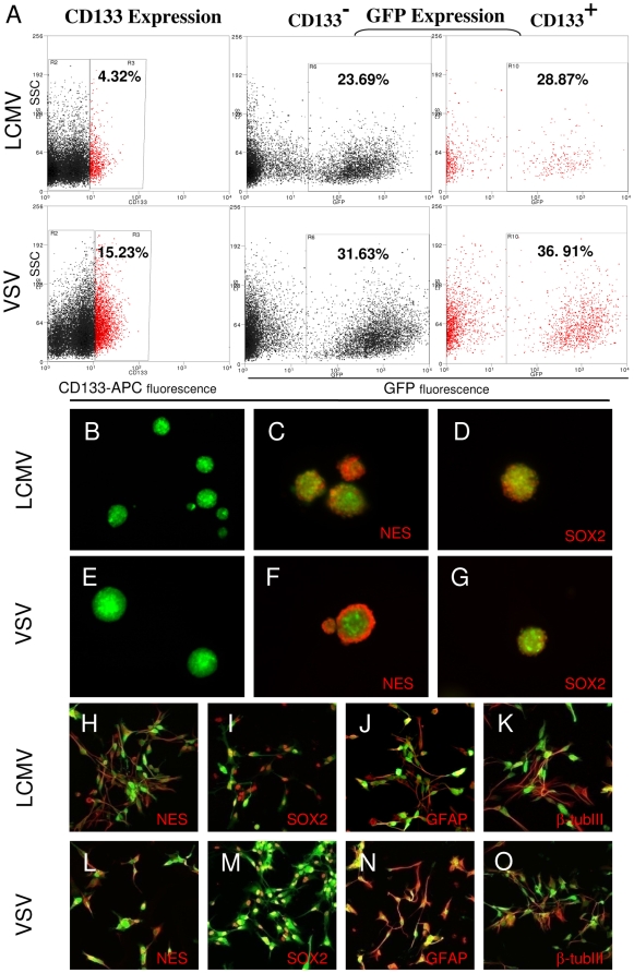
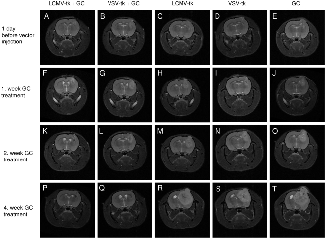
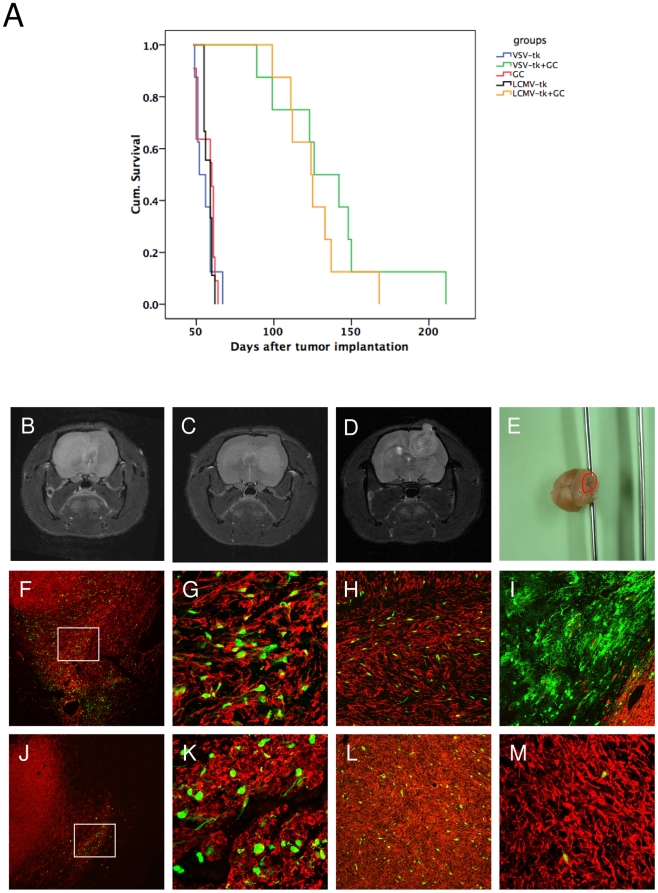
Methodology/principal findings: We used a rodent xenograft model that recapitulates the invasive and angiogenic features of human glioblastoma to analyze the transduction pattern and therapeutic efficacy of lentiviral pseudotyped vectors. Both, lymphocytic choriomeningitis virus glycoprotein (LCMV-GP) and vesicular stomatitis virus glycoprotein (VSV-G) pseudotyped lentiviral vectors very efficiently transduced human glioblastoma cells in vitro and in vivo. In contrast, pseudotyped gammaretroviral vectors, similar to those evaluated for clinical therapy of glioblastoma, showed inefficient gene transfer in vitro and in vivo. Both pseudotyped lentiviral vectors transduced cancer stem-like cells characterized by their CD133-, nestin- and SOX2-expression, the ability to form spheroids in neural stem cell medium and to express astrocytic and neuronal differentiation markers under serum conditions. In a therapeutic approach using the suicide gene herpes simplex virus thymidine kinase (HSV-1-tk) fused to eGFP, both lentiviral vectors mediated a complete remission of solid tumors as seen on MRI resulting in a highly significant survival benefit (p<0.001) compared to control groups. In all recurrent tumors, surviving eGFP-positive tumor cells were found, advocating prodrug application for several cycles to even enhance and prolong the therapeutic effect.

Conclusions/significance: In conclusion, lentiviral pseudotyped vectors are promising candidates for gene therapy of glioma in patients. The inefficient gene delivery by gammaretroviral vectors is in line with the results obtained in clinical therapy for GBM and thus confirms the high reproducibility of the invasive glioma animal model for translational research.


Conflict of interest statement

Competing Interests: The authors have declared that no competing interests exist.

Figures

Figure 1
Figure 1. Transduction of glioma spheroids by lenti- and retroviral vectors.
Human glioblastoma spheroids derived from a patient biopsy or after serial passaging in nude rats (high generation) were infected by lentiviral vectors pseudotyped with LCMV-GP (5×10⁴) or VSV-G (5×10⁴) or by retroviral vectors pseudotyped with LCMV-GP (1×10⁵). All vectors were expressing the marker gene eGFP. Spheroids were analyzed by confocal microscopy for eGFP expression 7 days after infection. Pictures show eGFP (green), phase contrast and an overlay of both. Low gen.: spheroids directly derived from patient material (low generation). High gen.: spheroids derived from serial in vivo passages in the rat brain (high generation). Original magnification 100×.
Figure 2
Figure 2. Glioblastoma patient biopsy and xenograft tumor show similar histopathology.
Paraffin sections of the patient tumor and the xenograft tumor were stained with H&E (A-D) or immunostained with human-specific anti-nestin antibodies (E,F). Patient tumor (A) and xenograft tumor (B) show angiogenic features of human glioblastoma with palisading necrosis (arrow) and vascular proliferates (arrowheads). Higher magnification of the patient (C) and xenograft tumor (D) demonstrate similar tumor cell morphology with polymorphic nuclei in the vicinity of a tumor necrosis. Patient (E) and xenograft tumor (F) show strong nestin expression of tumor cells. Single tumor cell infiltration into the white matter is observed in the xenograft tumor (F). The patient biopsy was derived only from the solid tumor core. A,B,E,F: Original magnification 100×. C,D: Original magnification 200×.
Figure 3
Figure 3. Lentiviral vectors efficiently transduce human glioma cells in vivo.
Intracranial gliomas were infected with LCMV-GP or VSV-G pseudotyped lentiviral vectors or with LCMV-GP pseudotyped retroviral vectors expressing eGFP 3–4 weeks after spheroid implantation and analyzed by fluorescence (A,D,G) or confocal scanning laser microscopy (B,C,E,F,H) 7 days after infection. The confocal images show overlay of eGFP (green fluorescence) and human-specific nestin (red fluorescence). Tumors were efficiently transduced by lentiviral LCMV-GP (A-C) and VSV-G (D-F) pseudotyped vectors, while retroviral vectors only transduced few scattered tumor cells (G,H; arrows). Transduced glioma cells expressed human-specific nestin in solid (B,E,H) and invasive tumor areas (C,F; arrows). (I) Transduction efficiency of lentiviral vectors was compared quantitatively by measuring the volume of transduction on histological sections using a fluorescence microscope and Nikon imaging software. LCMV transduced tumors (n = 3) showed a higher transduction volume than VSV transduced tumors (n = 3), however, the difference was not statistically significant (p = 0.269). A,D: Original magnification 40×. C,E,G;H: Original magnification 100×. D, F: Original magnification 200×.
Figure 4
Figure 4. Specificity of tumor cell transduction by lentiviral vectors.
Intracranial gliomas were infected with LCMV-GP- or VSV-G-pseudotyped lentiviral vectors expressing eGFP 3–4 weeks after tumor implantation and analyzed confocal laser scanning microscopy 7 days after infection. Transduction of invasive tumor cells was analyzed after staining with human-specific nestin antibodies. Host neurons and astrocytes were labeled using antibodies against NeuN and GFAP. Invasive areas showed single cell invasion by tumor cells (A-C, E, F) or a subependymal accumulation of tumor cells (D,G,H). LCMV pseudotyped vectors specifically transduced invasive glioma cells (A-C). VSV-G pseudotyped vectors showed specific transduction of tumor cells in some invasive areas (D-F), but also transduction of single normal brain cells in others (G,H; arrows). Transduced normal brain cells were mostly detected by morphologic criteria (more processes), as the staining (GFAP or NeuN) not always matched with the transduced cells. A,D: nestin staining, magnification 200×. B,E,G: GFAP staining, magnification 200×. C,F,H: NeuN staining, magnification 200×.
Figure 5
Figure 5. Lentiviral vectors transduce cancer stem-like cells.
Intracranial gliomas were infected with LCMV-GP or VSV-G pseudotyped lentiviral vectors expressing eGFP 3–4 weeks after tumor implantation. Tumors were excised when large lesions appeared on MRI and were enzymatically dissociated. The transduction of CD133 positive cells was measured by flow cytometry. (A) LCMV-GP and VSV-G pseudotyped vectors transduce CD133 positive and negative tumor cells. The fraction of transduced (GFP-positive) cells is slightly higher in CD133 positive cells (right column) compared to CD133 negative cells (middle column). GFP+ cells were sorted, cultured in the presence or absence of serum and analyzed by fluorescence (B-G) or confocal microscopy (H-O). LCMV-GP (B) and VSV-G (E) transduced cells form spheroids upon culture in serum-free neural basal medium supplemented with EGF and bFGF. Transduced spheroids express the neural stem cell markers nestin (C,F) and SOX2 (D,G). Transduced cells cultured in serum-containing medium express the stem cell markers nestin (H,L) and SOX2 (I,M), but also the differentiation markers GFAP (J,N) and beta-tubulinIII (K,O). The pictures C,D,E,F,H-O show overlay of the virus-delivered transgene (eGFP, green) and detected antigen (Alexa-647, red).
Figure 6
Figure 6. Tumors treated with lentiviral vectors and ganciclovir show complete remission on MRI.
Representative three-dimensional MRI (T2 RARE). (A,F,K,P) Lentiviral LCMV-GP vectors with ganciclovir treatment. (B,G,L,Q) Lentiviral VSV-G vectors with ganciclovir treatment. (C,H,M,R) Lentiviral LCMV-GP vectors without ganciclovir treatment. (D,I,N,S) Lentiviral VSV-G vectors without ganciclovir treatment. (E,J,O,T) ganciclovir treatment only. Time points after tumor implantation: (A-E) 1 day before vector injection. (F-J) 1. week ganciclovir treatment. (K-O) 2. week ganciclovir treatment. (P-T) 4. week ganciclovir treatment.
Figure 7
Figure 7. Therapeutic efficiency of LCMV-GP and VSV-G pseudotyped lentiviral vectors in vivo.
Intracranial gliomas were injected with LCMV-GP or VSV-G pseudotyped lentiviral vectors expressing HSV-1-tk fused to eGFP 3 weeks after tumor implantation. 7 days after vector infection, animals in both treated groups and in one control group were treated with ganciclovir for 30 days. (A) Kaplan-Meier survival curve. The survival benefit for both treatment groups compared to control groups was statistically significant (P<0.001; log-rank test). There was no significant difference in survival between the two treatment groups. (B-D) Representative MRI (T2 RARE) of recurrent tumors in the LCMV- (B,C) and VSV-treated (D,E) group. (B) Invasive contralateral recurrence. (C) Invasive local and contralateral recurrence. (D) More circumscribed local recurrence. (E) Macroscopic picture of a rat brain with a recurrence in the cerebellum (red circle), treated with VSV-G pseudotyped vectors and GC. (F-M) Sections of recurrent tumors were stained with antibodies against human-specific nestin and analyzed by fluorescence (F,J) or confocal microscopy (G-I,K-M). Pictures show overlay of nestin (red) and eGFP transgene (green). (F-I) Recurrent tumors of animals treated with VSV-G pseudotyped lentiviral vectors. (F) Recurrent tumor with GFP-positive cells in the invasive area. (G) Higher magnification of (F). (H) GFP-positive tumor cells in the corpus callosum region. (I) GFP-positive normal brain cells at the tumor border. (J-M) Recurrent tumors of animals treated with LCMV-GP pseudotyped lentiviral vectors. (J) GFP-positive tumor cells in residual small lesion from the primary tumor. The recurrent tumor is growing from the contralateral hemisphere over the corpus callosum to the ipsilateral hemisphere (arrows). (K) Higher magnification of (J). (L) GFP-positive tumor cells in the solid ipsilateral recurrent lesion. (M) Few GFP-positive cells in a contralateral recurrent tumor. F,J: Magnification 40×. G,K,M: Magnification 200×. H,I,L: Magnification 100×.

References

    1. Stupp R, Mason WP, van den Bent MJ, Weller M, Fisher B, et al. Radiotherapy plus concomitant and adjuvant temozolomide for glioblastoma. N Engl J Med. 2005;352:987–996. - PubMed
    1. Mahesparan R, Read TA, Lund-Johansen M, Skaftnesmo KO, Bjerkvig R, et al. Expression of extracellular matrix components in a highly infiltrative in vivo glioma model. Acta Neuropathol. 2003;105:49–57. - PubMed
    1. Sakariassen PO, Prestegarden L, Wang J, Skaftnesmo KO, Mahesparan R, et al. Angiogenesis-independent tumor growth mediated by stem-like cancer cells. Proc Natl Acad Sci U S A. 2006;103:16466–16471. - PMC - PubMed
    1. Huszthy PC, Goplen D, Thorsen F, Immervoll H, Wang J, et al. Oncolytic herpes simplex virus type-1 therapy in a highly infiltrative animal model of human glioblastoma. Clin Cancer Res. 2008;14:1571–1580. - PubMed
    1. Mineta T, Rabkin SD, Yazaki T, Hunter WD, Martuza RL. Attenuated multi-mutated herpes simplex virus-1 for the treatment of malignant gliomas. Nat Med. 1995;1:938–943. - PubMed

Publication types