. 2009 Aug;238(8):2095-102.
doi: 10.1002/dvdy.22021.

Regional expression of MTG genes in the developing mouse central nervous system

Affiliations

Regional expression of MTG genes in the developing mouse central nervous system

Amin Alishahi et al. Dev Dyn. 2009 Aug.

Abstract

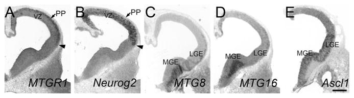
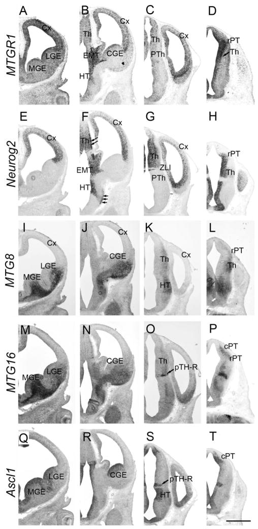
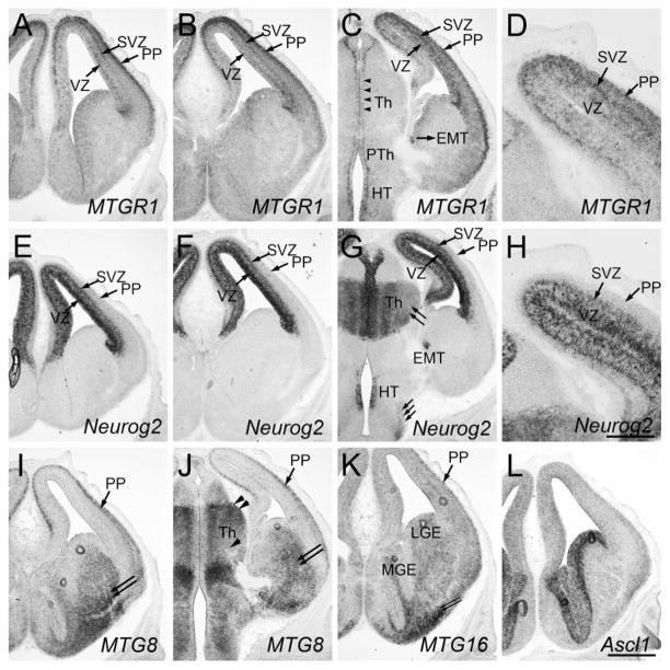
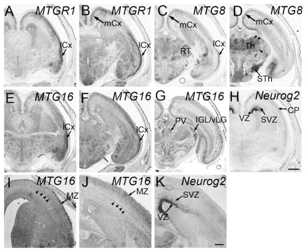
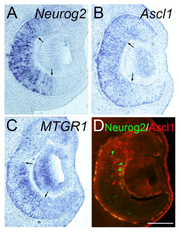
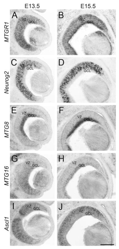
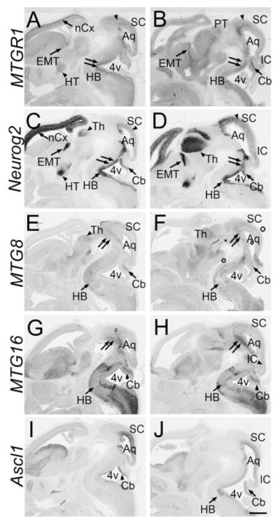
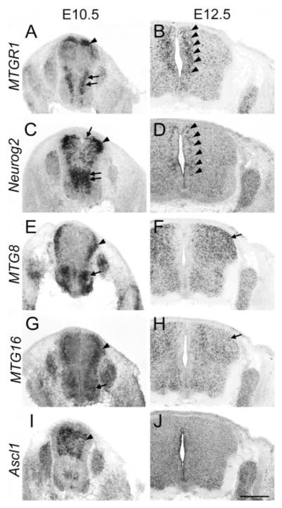
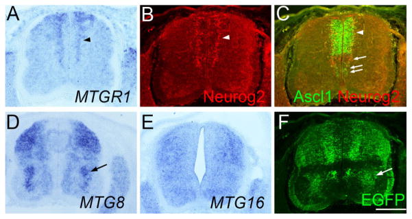
Myeloid translocation gene (MTG) proteins are transcriptional repressors that are highly conserved across species. We studied the expression of three members of this gene family, MTGR1, MTG8, and MTG16 in developing mouse central nervous system by in situ hybridization. All of these genes are detected as early as embryonic day 11.5. Because these genes are known to be induced by proneural genes during neurogenesis, we analyzed the expression of MTG genes in relation to two proneural genes, Neurog2 (also known as Ngn2 or Neurogenin 2) and Ascl1 (also known as Mash1). While MTGR1 are generally expressed in regions that also express Neurog2, MTG8 and MTG16 expression is associated more tightly with that of Ascl1-expressing neural progenitor cells. These results suggest the possibility that expression of MTG genes is differentially controlled by specific proneural genes during neurogenesis.


Figures

Fig. 1
Fig. 1
Telencephalon at E10.5. Adjacent frontal sections are shown. Midline is to the left. In situ hybridization for MTGR1 (A), Neurog2 (B), MTG8 (C), MTG16 (D), and Ascl1 (E) is shown. PP, preplate; VZ, ventricular zone. Scale bar = 200 μm.
Fig. 2
Fig. 2
Forebrain at E11.5. Frontal sections are shown. Sections in the same column are adjacent to each other. Left columns are more frontal than right columns. Midline is to the left. In situ hybridization for MTGR1 (A–D), Neurog2 (E–H), MTG8 (I–L), MTG16 (M–P), and Ascl1 (Q–T) is shown. Cx, cortex; MGE, medial ganglionic eminence; LGE, lateral ganglionic eminence; CGE, caudal ganglionic eminence; EMT, eminentia thalami; HT, hypothalamus; rPT, rostral pretectum; cPT, caudal pretectum; Th, thalamus; pTH-R, rostral thalamic progenitor domain; ZLI, zona limitans intrathalamica. Scale bar = 500 μm.
Fig. 3
Fig. 3
Forebrain at E13.5. Frontal sections are shown. A and E, B and F, C and G are adjacent sections. D and H are high-magnification views of C and G, respectively. Midline is to the left. In situ hybridization for MTGR1 (A–D), Neurog2 (E–H), MTG8 (I,J), MTG16 (K), and Ascl1 (L) is shown. PP, preplate; SVZ, subventricular zone; VZ, ventricular zone; MGE, medial ganglionic eminence; LGE, lateral ganglionic eminence; Th, thalamus; EMT, eminentia thalami; HT, hypothalamus. Scale bar = 200 μm for D,H and 500 μm for other panels.
Fig. 4
Fig. 4
Forebrain at E14.5 and E15.5. Frontal sections are shown. A–H, J,K are from E15.5, and I is from an E14.5 embryo. Midline is to the left. In situ hybridization for MTGR1 (A,B), MTG8 (C,D), MTG16 (E–G, I,J), Neurog2 (H,K). lCx, lateral cortex; mCx, medial cortex; RT, reticular nucleus; Th, thalamus; STh, subthalamic nucleus; PV, paraventricular nucleus of the thalamus; IGL/vLG, inter-geniculate leaflet/ventral lateral geniculate nucleus; VZ, ventricular zone; SVZ, subventricular zone. Scale bar = 500 μm for A–H, 200 μm for I–K.
Fig. 5
Fig. 5
Retina at E11.5. Frontal sections of the retina at E11.5. Midline is to the left. In situ hybridization for Neurog2 (A), Ascl1 (B), MTGR1 (C) as well as double immunohistochemistry of Ascl1 and Neurog2 (D) is shown. Red and green arrowheads in D indicate Neurog2-positive and Ascl1-positive cells, respectively. These two populations do not significantly overlap at this stage. Scale bar = 200 μm.
Fig. 6
Fig. 6
Retina at E13.5 and E15.5. Frontal sections of the forebrain at E13.5 (A,C,E,G,I) and E15.5 (B,D,F,H,J). Midline is to the left. In situ hybridization for MTGR1 (A,B), Neurog2 (C,D), MTG8 (E,F), MTG16 (G,H), and Ascl1 (I,J) is shown. VZ, ventricular zone; GCL, ganglion cell layer. Scale bar = 200 μm.
Fig. 7
Fig. 7
Sagittal sections at E12.5. Sagittal sections of the brain at E12.5. Front is to the left. In situ hybridization for MTGR1 (A,B), Neurog2 (C,D), MTG8 (E,F), MTG16 (G,H), and Ascl1 (I,J) is shown. nCx, neocortex; Th, thalamus; EMT, eminentia thalami; HT, hypothalamus; PT, pretectum; HB, hindbrain; Aq, cerebral aquaduct; 4v, fourth ventricle; SC, superior colliculus; IC, inferior colliculus; Cb, cerebellum. Scale bar = 500 μm.
Fig. 8
Fig. 8
Spinal cord at E10.5 and E12.5. Transverse sections of the spinal cord at the thoracic level at E10.5 (A,C,E,G,I) and E12.5 (B,D,F,H,J). In situ hybridization for MTGR1 (A,B), Neurog2 (C,D), MTG8 (E,F), MTG16 (G,H), and Ascl1 (I,J) is shown. Scale bar = 500 μm.
Fig. 9
Fig. 9
Analysis of Ascl1-BAC-EGFP embryos at E11.5. Transverse sections of the lumber spinal cord (A–F) of the same Ascl1-BAC-EGFP embryo at E11.5. Sections in A–F are adjacent to each other and B and C are from the same section. In situ hybridization for MTGR1 (A), MTG8 (D), MTG16 (E), as well as immunostaining for Neurog2 (B), Ascl1/Neurog2 (C), and EGFP (F) is shown. Scale bar = 200 μm.

References

    1. Amann JM, Chyla BJI, Ellis TC, Martinez A, Moore AC, Franklin JL, McGhee L, Meyers S, Ohm JE, Luce KS, Ouelette AJ, Washington MK, Thompson MA, King D, Gautam S, Coffey RJ, White-head RH, Hiebert SW. Mtgr1 is a transcriptional corepressor that is required for maintenance of the secretory cell lineage in the small intestine. Mol Cell Biol. 2005;25:9576 –9585. - PMC - PubMed
    1. Butt SJ, Fuccillo M, Nery S, Noctor S, Kriegstein A, Corbin JG, Fishell G. The temporal and spatial origins of cortical interneurons predict their physiological subtype. Neuron. 2005;48:591– 604. - PubMed
    1. Calabi F, Pannell R, Pavloska G. Gene targeting reveals a crucial role for MTG8 in the gut. Mol Cell Bio. 2001;21:5658 –5666. - PMC - PubMed
    1. Cao Y, Zhao H, Grunz H. XETOR regulates the size of the proneural domain during primary neurogenesis in Xenopus laevis. Mech Dev. 2002;119:35– 44. - PubMed
    1. Davis JN, McGhee L, Meyers S. The ETO (MTG8) gene family. Gene. 2003;303:1–10. - PubMed

Publication types

MeSH terms