. 2009 Aug 13;52(15):4694-715.
doi: 10.1021/jm900259h.

Discovery of leukotriene A4 hydrolase inhibitors using metabolomics biased fragment crystallography

Affiliations

Discovery of leukotriene A4 hydrolase inhibitors using metabolomics biased fragment crystallography

Douglas R Davies et al. J Med Chem. .

Erratum in

  • J Med Chem. 2010 Mar 11;53(5):2330-1

Abstract

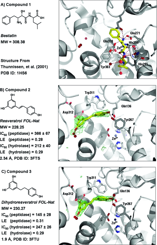
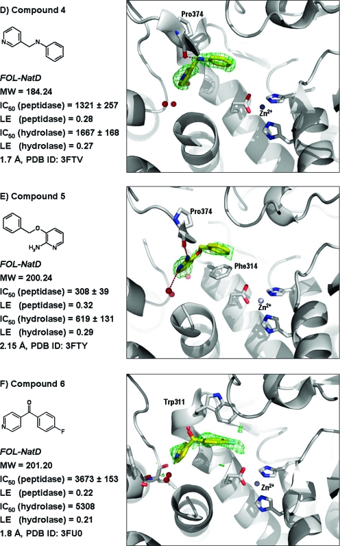
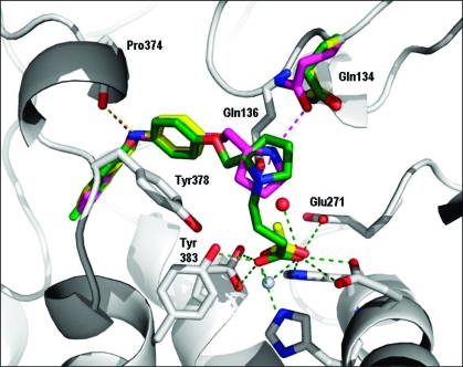
We describe a novel fragment library termed fragments of life (FOL) for structure-based drug discovery. The FOL library includes natural small molecules of life, derivatives thereof, and biaryl protein architecture mimetics. The choice of fragments facilitates the interrogation of protein active sites, allosteric binding sites, and protein-protein interaction surfaces for fragment binding. We screened the FOL library against leukotriene A4 hydrolase (LTA4H) by X-ray crystallography. A diverse set of fragments including derivatives of resveratrol, nicotinamide, and indole were identified as efficient ligands for LTA4H. These fragments were elaborated in a small number of synthetic cycles into potent inhibitors of LTA4H representing multiple novel chemotypes for modulating leukotriene biosynthesis. Analysis of the fragment-bound structures also showed that the fragments comprehensively recapitulated key chemical features and binding modes of several reported LTA4H inhibitors.


Figures

Figure 1
Figure 1
Conceptual organization of key components of the fragments of life library, with example fragments. The current FOL library (1329 molecules) contains chemically tractable natural small molecule metabolites such as bestatin 1 (FOL-Nat), metabolite-like compounds and their bioisosteres (FOL-NatD), and protein architecture mimetics (FOL-Biaryl). Any natural molecule with MW < 350 Da that exists as a substrate, natural product, or allosteric regulator of any metabolic pathway in any cell type is a potential candidate FOL-Nat fragment. Example fragments 11 and 13 that contain the privileged scaffold indole(98) from the serotonin and auxin biosynthetic pathways are shown. FOL-NatD fragments are defined as heteroatom containing derivatives, analogues, or fragments of any FOL-Nat molecule. FOL-Biaryl fragments were selected from a variety of biaryl compounds that are potential mimics of protein α, β, or γ turns. At the right-hand side of the figure, compound 10 can be seen superimposed on a β-turn of a protein structure (residues Ala20-Ala21-Asp22-Ser23 of PDB ID 1RTP). An additional FOL-Biaryl component is also shown, superimposed on an α-turn (residues Ser65-Ile66-Leu67-Lys68 of PDB ID 1RTP).
Figure 2
Figure 2
Panels showing ligand binding to LTA4H for compounds described in the manuscript. Enzyme assay IC50 values are in μM, human whole blood cell assay IC50 values are in nM when reported, and ligand efficiency (LE) values are in kcal/(mol·heavy atom). Compound structures are displayed as yellow stick structures, and LTA4H is displayed in gray. Green mesh corresponds to the FoFc (difference) electron density at the 3.0σ level of the crystal structure with the compound omitted from the model. Polar contacts with LTA4H and/or bound water molecules are shown as red dashed lines. PDB IDs for each structure are indicated.
Figure 2
Figure 2
Panels showing ligand binding to LTA4H for compounds described in the manuscript. Enzyme assay IC50 values are in μM, human whole blood cell assay IC50 values are in nM when reported, and ligand efficiency (LE) values are in kcal/(mol·heavy atom). Compound structures are displayed as yellow stick structures, and LTA4H is displayed in gray. Green mesh corresponds to the FoFc (difference) electron density at the 3.0σ level of the crystal structure with the compound omitted from the model. Polar contacts with LTA4H and/or bound water molecules are shown as red dashed lines. PDB IDs for each structure are indicated.
Figure 3
Figure 3
Resveratrol structures. (A) The structure of resveratrol (2) (yellow) is superimposed upon dihydroresveratrol (3) (cyan). (B) Superposition of the structure of dihydroresveratrol (3) is shown in the absence (cyan) and presence (magenta) of bestatin. Structurally conserved water molecules and the side chain of residue Phe362 are also color-coded to show the differences in the structures with and without bestatin. For crystallographic data, see Supporting Information Table 1.
Scheme 1
Scheme 1. Synthetic Routes for New Molecules Inspired by the LTA4H Binding Modes of Compounds 4, 6, and 7
Reagents and conditions: (a) AlCl3, 10 °C, nitrobenzene, 90%; (b), BBr3, CH2Cl2, −78 °C → 0 °C, 85%; (c) NaH/DMF, 0 °C → rt → 90 °C; (d) 1 M HCl/Et2O, 80−97%; (e) thiophene-3-boronic acid (2 equiv), Pd(OAc)2 (25 mol %), K2CO3/EtOH/DME, 90 °C, 60%.
Figure 4
Figure 4
Elaboration of 4. Refined crystal structures of LTA4H in complex with molecules in the compound 4 elaboration series are shown. Compound 4 and acetate from the “fragment plus fragment” structure are displayed as yellow stick structures, while compound 14 is magenta and compound 19 (DG-051) is green. The side chain conformations of LTA4H bound to each of these compounds are essentially superimposable (gray side chains) with the exception of Gln134, which demonstrates some flexibility in its conformation as represented by DG-051 (green), compound 4 and acetate (yellow), and compound 14 (magenta). Polar contacts are displayed as color-coded dashed lines.
Figure 5
Figure 5
Elaboration of 6. (A) Superposition of compounds 6 (yellow), 17 (green), and 18 (cyan). Invariant portions of the LTA4H structure are colored gray, and compound-specific polar contacts, side chain conformations, and bound water molecules are color-coded by compound. (B) Superpostion of 18 (cyan) with the thiazole-containing fragment 7 (rose) and the pyrrolidine containing compound 14 (magenta). Both 7 and 18 displace the central structurally conserved water molecule, and both 14 and 18 have pyrrolidine moieties that occupy nearly the same space in the LTA4H structures.
Scheme 2
Scheme 2. Elaboration of Fragment 6
Reagents and conditions used are as follows: (a) 1-(2-hydroxyethyl)pyrrolidine, K-OtBu, DMSO, 90 °C, 16 h, 54%; (b) NaBH4, MeOH, 40 °C, 4 h.
Figure 6
Figure 6
Elaboration of 5-hydroxyindole 11, showing superposition of the indole-containing fragment structures: 5-hydroxyindole 11 (dark green), 5-chlororindole 12 (magenta), 5-fluoroindole 13 (cyan), and derivative compounds 15 (yellow) and 16 (pink).
Scheme 3
Scheme 3. Elaboration of Fragment 11
Reagents and conditions: (a) 1-(2-chloroethyl)pyrrolidine, K2CO3, acetone, 19%; (b) 1-bromo-2-chloroethane, K2CO3, 2-butanone, reflux 60 h, 17%; (c) piperidine-4-carboxylic acid ethyl ester, HCl (2 equiv), KI (0.3 equiv), K2CO3, DMF, rt → 90 °C, 57%; (d) aq NaOH, EtOH, rt.

Similar articles

Cited by

References

    1. Zheng H.; Chruszcz M.; Lasota P.; Lebioda L.; Minor W. Data mining of metal ion environments present in protein structures. J. Inorg. Biochem. 2008, 102, 1765–1776. - PMC - PubMed
    1. Tan X.; Calderon-Villalobos L. I.; Sharon M.; Zheng C.; Robinson C. V.; Estelle M.; Zheng N. Mechanism of auxin perception by the TIR1 ubiquitin ligase. Nature 2007, 446, 640–645. - PubMed
    1. Shimada A.; Ueguchi-Tanaka M.; Nakatsu T.; Nakajima M.; Naoe Y.; Ohmiya H.; Kato H.; Matsuoka M. Structural basis for gibberellin recognition by its receptor GID1. Nature 2008, 456, 520–523. - PubMed
    1. Kuntz I. D.; Chen K.; Sharp K. A.; Kollman P. A. The maximal affinity of ligands. Proc. Natl. Acad. Sci. U.S.A. 1999, 96, 9997–10002. - PMC - PubMed
    1. Wishart D. S.; Knox C.; Guo A. C.; Eisner R.; Young N.; Gautam B.; Hau D. D.; Psychogios N.; Dong E.; Bouatra S.; Mandal R.; Sinelnikov I.; Xia J.; Jia L.; Cruz J. A.; Lim E.; Sobsey C. A.; Shrivastava S.; Huang P.; Liu P.; Fang L.; Peng J.; Fradette R.; Cheng D.; Tzur D.; Clements M.; Lewis A.; De Souza A.; Zuniga A.; Dawe M.; Xiong Y.; Clive D.; Greiner R.; Nazyrova A.; Shaykhutdinov R.; Li L.; Vogel H. J.; Forsythe I. HMDB: a knowledgebase for the human metabolome. Nucleic Acids Res. 2008, 37 (Database Issue), D603–D610. - PMC - PubMed

Publication types

LinkOut - more resources