Comparative Study
. 2009 Aug 25;48(33):8032-43.
doi: 10.1021/bi9010013.

The amino acid motif L/IIxxFE defines a novel actin-binding sequence in PDZ-RhoGEF

Affiliations
Comparative Study

The amino acid motif L/IIxxFE defines a novel actin-binding sequence in PDZ-RhoGEF

Jayashree Banerjee et al. Biochemistry. .

Abstract

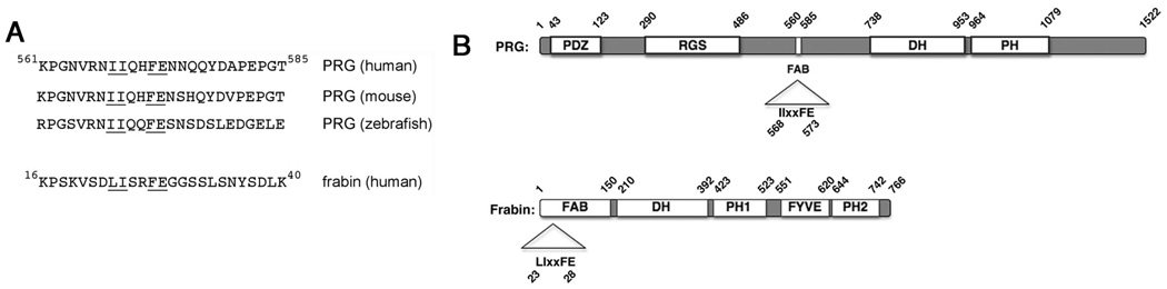
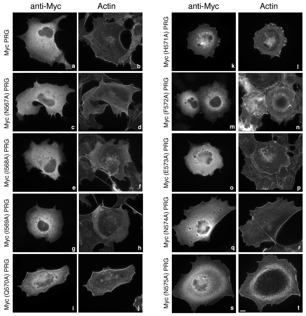
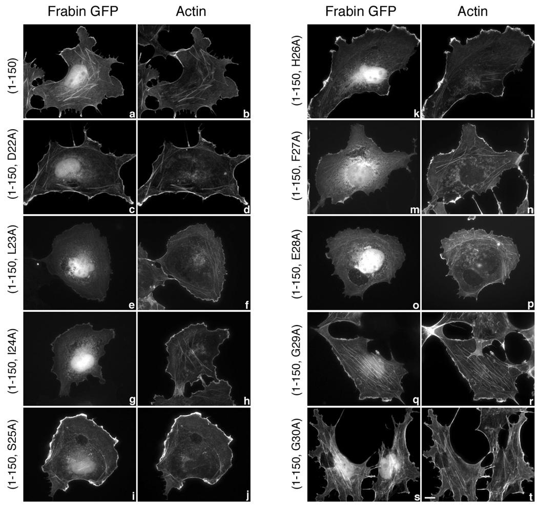
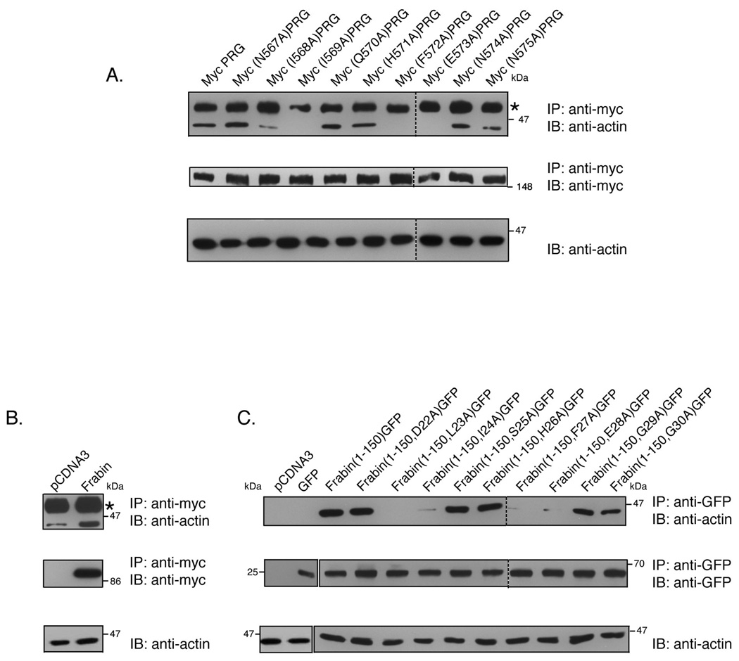
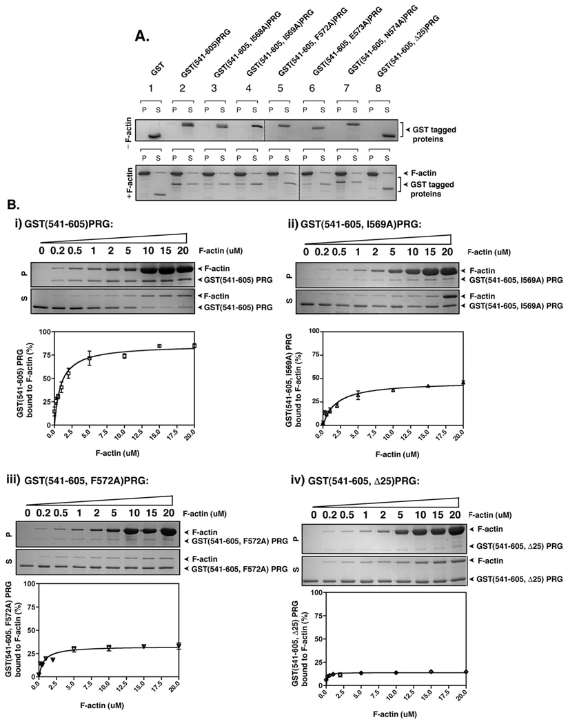
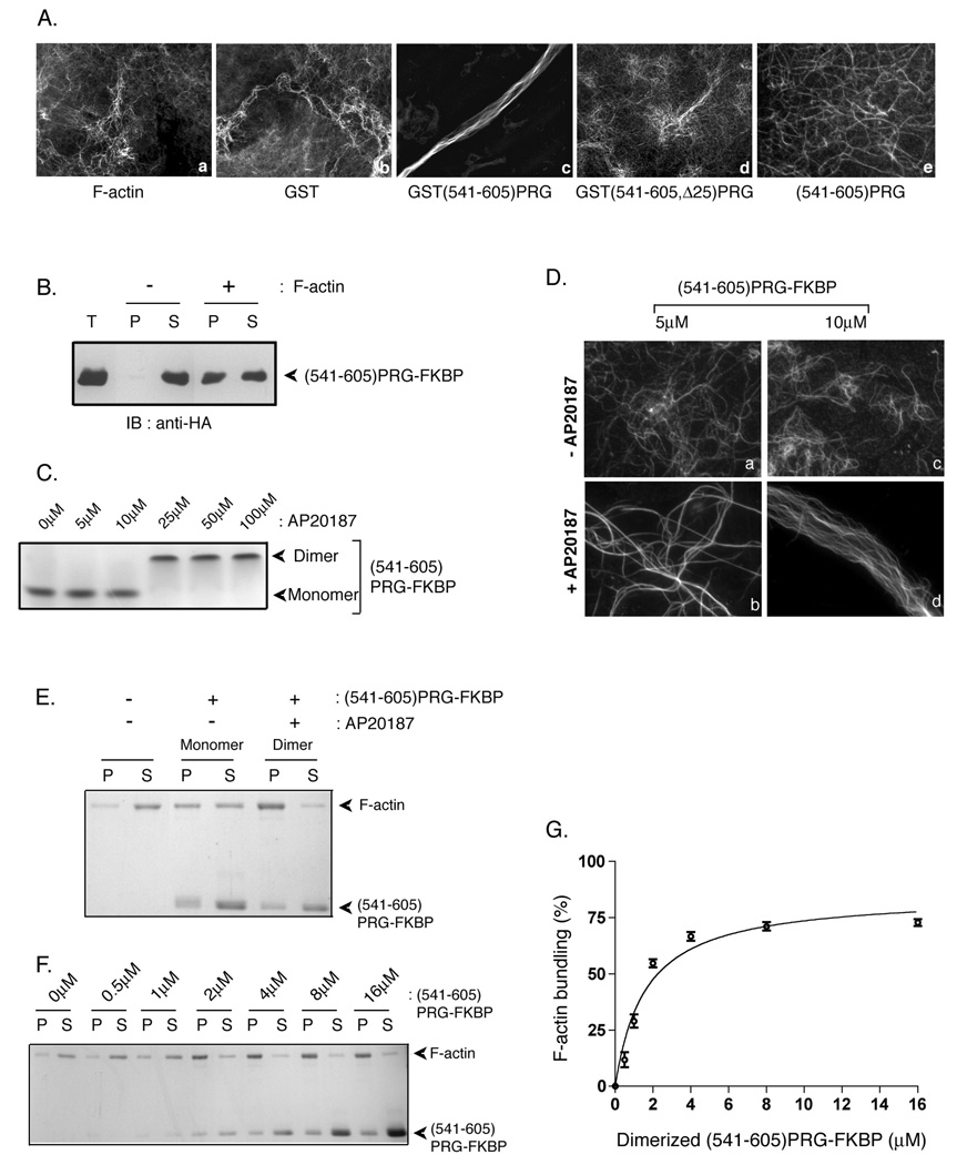
PDZ-RhoGEF is a member of the regulator family of G protein signaling (RGS) domain-containing RhoGEFs (RGS-RhoGEFs) that link activated heterotrimeric G protein alpha subunits of the G12 family to activation of the small GTPase RhoA. Unique among the RGS-RhoGEFs, PDZ-RhoGEF contains a short sequence that localizes the protein to the actin cytoskeleton. In this report, we demonstrate that the actin-binding domain, located between amino acids 561 and 585, directly binds to F-actin in vitro. Extensive mutagenesis identifies isoleucine 568, isoleucine 569, phenylalanine 572, and glutamic acid 573 as being necessary for binding to actin and for colocalization with the actin cytoskeleton in cells. These results define a novel actin-binding sequence in PDZ-RhoGEF with a critical amino acid motif of IIxxFE. Moreover, sequence analysis identifies a similar actin-binding motif in the N-terminus of the RhoGEF frabin, and as with PDZ-RhoGEF, mutagenesis and actin interaction experiments demonstrate an LIxxFE motif, consisting of the key amino acids leucine 23, isoleucine 24, phenylalanine 27, and glutamic acid 28. Taken together, results with PDZ-RhoGEF and frabin identify a novel actin-binding sequence. Lastly, inducible dimerization of the actin-binding region of PDZ-RhoGEF revealed a dimerization-dependent actin bundling activity in vitro. PDZ-RhoGEF exists in cells as a dimer, raising the possibility that PDZ-RhoGEF could influence actin structure in a manner independent of its ability to activate RhoA.


Figures

Figure 1
Figure 1. Actin-binding motif of PRG and frabin
(A) The amino acid sequence encompassing the predicted actin-binding motif in human PRG (amino acids 561–585) is shown along with sequences in similar locations in the mouse and zebrafish PRG orthologs. In addition, the predicted actin-binding motif (amino acids 16–40) of frabin is shown. The underlined amino acids indicate similarities among PRG orthologs and frabin, and the underlined amino acids represent a minimal actin-binding motif, L/IIxxFE, defined in this report. (B) The locations of the actin-binding motif in the overall structure of PRG and frabin are indicated.
Figure 2
Figure 2. Subcellular localization of PRG and mutants
COS-7 cells were transfected with 1 µg of an expression vector encoding Myc epitope–tagged PRG (a and b), Myc(N567A)PRG (c and d), Myc(I568A)PRG (e and f), Myc(I569A)PRG (g and h), Myc(Q570A)PRG (i and j), Myc(H571A)PRG (k and l), Myc(F572A)PRG (m and n), Myc(E573A)PRG (o and p), Myc(N574A)PRG (q and r) or Myc(N575A)PRG (s and t). 24 h post-transfection, cells were fixed and subjected to immunofluorescence microscopy as described in “Materials and Methods”. Expressed proteins were detected with an anti-Myc 9E10 antibody (a, c, e, g, I, k, m, o, q and s) followed by Alexa 594 conjugated to an anti-mouse antibody. Actin was visualized in the same cells by co-staining with Alexa 488 conjugated to phalloidin (b, d, f, h, j, l, n, p, r and t). Bar, 10 µm. Images shown are single cells representative of at least five separate experiments in which more than fifty cells were viewed in each experiment. Note that wild type PRG (a) and unaffected PRG mutants (c, i, k, q, s) show a modest co-localization with F-actin at the cell periphery, whereas I568A, I569A, F572A, and E573A mutants of PRG (e, g, m, o) reproducibly display a complete loss of staining at the cell periphery.
Figure 3
Figure 3. Subcellular localization of frabin mutants
COS-7 cells were transiently transfected with 1 µg expression vectors encoding GFP-tagged frabin mutants, frabin(1–150) (a and b), frabin(1–150, D22A) (c and d), frabin(1–150, L23A) (e and f), frabin(1–150, I24A) (g and h), frabin(1–150, S25A) (i and j), frabin(1–150, H26A) (k and l), frabin(1–150, F27A) (m and n), frabin(1–150, E28A) (o and p), frabin(1–150, G29A) (q and r) and frabin(1–150, G30A) (s and t). Expressed proteins were visualized by GFP fluorescence (a, c, e, g, I, k, m, o, q and s), and actin was visualized (b, d, f, h, j, l, n, p, r and t) in the same cells by staining with Alexa 594 conjugated to phalloidin. Bar, 10 µm. Images shown are single cells representative of at least five separate experiments in which more than fifty cells were viewed in each experiment. Note that frabin(1–150) (a) and unaffected frabin(1–150) mutants (c, i, k, q, s) show an intense GFP signal at the cell periphery and at intracellular stress fibers that co-localizes with F-actin, whereas L23A, I24A, F27A, and E28A frabin(1–150) (e, g, m, o) reproducibly display a weak GFP signal at the cell periphery. The weak signal at the cell periphery observed with the four frabin mutants (e, g, m, o) is identical to the background signal of GFP alone.
Figure 4
Figure 4. PRG and frabin co-immunoprecipitate with actin
(A) COS-7 cells were transiently transfected with 7 µg of an expression vector encoding PRG or the indicated mutants. PRG was immunoprecipitated from cell lysates with a mouse monoclonal anti-Myc antibody, and immunoprecipitates were analyzed by immunoblotting using anti-actin mAb (upper panel). Immunoprecipitation of the Myc-tagged PRG proteins was confirmed by immunoblotting of the immunoprecipitates using anti-Myc antibody (middle panel). Presence of actin in the lysates was detected by immunoblotting using anti-actin mAb (lower panel). (B) COS-7 cells were transfected with 7 µg empty vector or with expression plasmid for Myc-tagged full-length frabin. Cells were lysed and lysates were subjected to immunoprecipitation by monoclonal anti-Myc antibody. Immunoprecipitates were analyzed by immunoblot using anti-actin mAb (upper panel) and anti-Myc Ab (middle panel). Actin in the cell lysates was detected using anti-actin mAb (lower panel). (C) COS-7 cells were transiently transfected with 7 µg of empty vector, GFP alone or with the indicated GFP-tagged frabin mutants. Cell lysates were immunoprecipitated with polyclonal anti-GFP antibody and immunoprecipitates were probed for actin using anti-actin mAb (upper panel). Immunoprecipitation of GFP-tagged frabin constructs were confirmed by immunoblot of immunoprecipitates using anti-GFP antibody (middle panel), and presence of actin in the cell lysates was confirmed by anti-actin mAb (lower panel). Asterisks (*) indicate the antibody heavy chain that migrates slower than actin.
Figure 5
Figure 5. PRG binds to F-actin in vitro
(A) 2 µg of the indicated recombinant proteins was incubated with or without 40 µg of freshly polymerized F-actin at room temperature for 30 min and then centrifuged at 160,000g for 90 min. Equal aliquots of resuspended pellet (P) and supernatant (S) were analyzed by SDS-PAGE and colloidal blue staining. (B) Quantification of F-actin binding activity of recombinant PRG mutants, GST(541–605)PRG (i), GST(541–605, I569A)PRG (ii), GST(541–605, F572A)PRG and (iv) GST(541–605, Δ25)PRG. 3 µM of the indicated mutants of PRG were incubated with variable concentrations F-actin (0.2 – 20 µM) and co-sedimented at 160,000g for 90 min. Equal aliquots of resuspended pellet (P) and supernatant (S) were analyzed by SDS-PAGE and colloidal blue staining (upper panels), and the bands were quantified by densitometry. The percentage of F-actin bound PRG mutants was calculated as the percentage recovered in the pellet (P) over total protein (S+P). The graphs represent the means ±S.D. of three independent experiments.
Figure 6
Figure 6. PRG induces F-actin bundling in vitro
(A) F-actin (1.2 µM) was incubated alone (a) or with 7 µM GST (b), GST(541–605)PRG (c), GST(541–605, Δ25)PRG (d), or (541–605)PRG in which GST was removed by proteolysis (e) for 30 min on ice. After staining with phalloidin, actin filaments and bundles were observed by fluorescence microscopy. (B) (541–605)PRG-FKBP (GST was removed by proteolysis) incubated with or without freshly polymerized actin was subjected to high speed co-sedimentation assay at 160,000g for 90 min. Equal aliquots of pellet and supernatant fractions along with total protein (T) were immunoblotted using anti-HA antibody to detect (541–605)PRG-FKBP. (C) 25 µM (541–605)PRG-FKBP was incubated with variable concentrations (5–100 µM) of AP20187 for 15 min at room temperature. Samples were subject to native gel electrophoresis followed by colloidal blue staining. 25 µM (541–605)PRG-FKBP was used to observe consistent colloidal blue staining. (D) (541–605)PRG-FKBP (5 µM and 10 µM) treated without (a and c) and with (b and d) AP20187 was incubated with 1.2 µM freshly polymerized F-actin. Following incubation, F-actin was stained with phalloidin and observed by fluorescence microscopy. (E) 2 µM monomeric or dimeric (541–605)PRG-FKBP was incubated with 1 µM freshly polymerized F-actin, and samples were centrifuged at 10,000g for 30 min. Equal amounts of pellet (P) and supernatant (S) fractions were resolved by SDS-PAGE and stained using colloidal blue. (F) To quantitate F-actin bundling activity of dimerized (541–605)PRG-FKBP, 0, 0.5, 1, 2, 4, 8 or 16 µM of dimerized (541–605)PRG-FKBP was incubated with 1 µM of F-actin. Samples were then centrifuged at 10,000g. amounts of pellet (P) and supernatant (S) fractions were run on SDS-PAGE and stained with colloidal blue. (G) F-actin in the 10,000g pellet fraction in (F) was quantitated using densitometry. The percentage of F-actin bundled using different amounts of dimerized (541–605)PRG-FKBP were calculated as the percentage recovered in the pellet (P) over total protein (S+P).The data represent the means ± S.D. of three independent experiments.

Similar articles

Cited by

References

    1. Wennerberg K, Rossman KL, Der CJ. The Ras superfamily at a glance. J Cell Sci. 2005;118:843–846. - PubMed
    1. Bustelo XR, Sauzeau V, Berenjeno IM. GTP-binding proteins of the Rho/Rac family: regulation, effectors and functions in vivo. Bioessays. 2007;29:356–370. - PMC - PubMed
    1. Ridley AJ, Hall A. The small GTP-binding protein rho regulates the assembly of focal adhesions and actin stress fibers in response to growth factors. Cell. 1992;70:389–399. - PubMed
    1. Ridley AJ, Paterson HF, Johnston CL, Diekmann D, Hall A. The small GTP-binding protein rac regulates growth factor-induced membrane ruffling. Cell. 1992;70:401–410. - PubMed
    1. Nobes CD, Hall A. Rho, rac, and cdc42 GTPases regulate the assembly of multimolecular focal complexes associated with actin stress fibers,lamellipodia, and filopodia. Cell. 1995;81:53–62. - PubMed

Publication types

MeSH terms