

Fluorescent glycosylamides produced by microscale derivatization of free glycans for natural glycan microarrays
- PMID: 19618966
- PMCID: PMC2746876
- DOI: 10.1021/cb900067h
Fluorescent glycosylamides produced by microscale derivatization of free glycans for natural glycan microarrays
Abstract
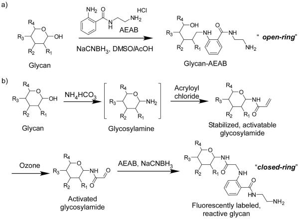
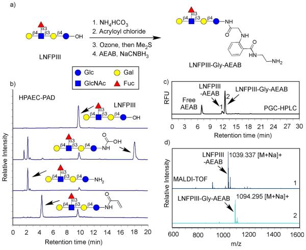
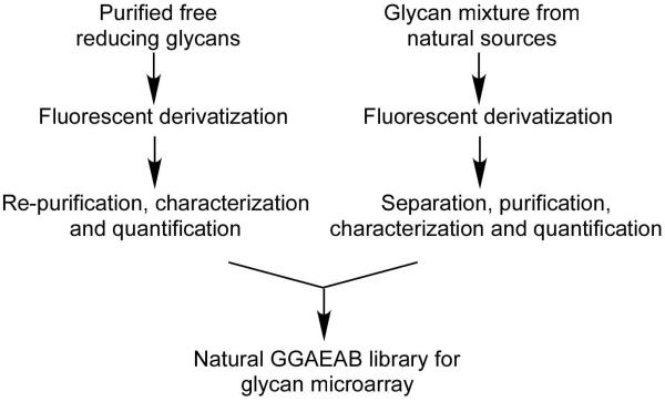
A novel strategy for creating naturally derived glycan microarrays has been developed. Glycosylamines are prepared from free reducing glycans and stabilized by reaction with acryloyl chloride to generate a glycosylamide in which the reducing monosaccharide has a closed-ring structure. Ozonolysis of the protected glycan yields an active aldehyde, to which a bifunctional fluorescent linker is coupled by reductive amination. The fluorescent derivatives are easily coupled through a residual primary alkylamine to generate glycan microarrays. This strategy preserves structural features of glycans required for antibody recognition and allows development of natural arrays of fluorescent glycans in which the cyclic pyranose structure of the reducing-end sugar residue is retained.
Figures
Similar articles
-
Fluorescent glycan derivatives: their use for natural glycan microarrays.ACS Chem Biol. 2009 Sep 18;4(9):699-701. doi: 10.1021/cb9002078. ACS Chem Biol. 2009. PMID: 19761252
-
Novel cleavage of reductively aminated glycan-tags by N-bromosuccinimide to regenerate free, reducing glycans.ACS Chem Biol. 2013 Nov 15;8(11):2478-83. doi: 10.1021/cb400513k. Epub 2013 Sep 13. ACS Chem Biol. 2013. PMID: 23992636 Free PMC article.
-
Biotinyl-l-3-(2-naphthyl)-alanine hydrazide derivatives of N-glycans: versatile solid-phase probes for carbohydrate-recognition studies.Glycobiology. 1998 Mar;8(3):227-36. doi: 10.1093/glycob/8.3.227. Glycobiology. 1998. PMID: 9451032
-
Use of glycan microarrays to explore specificity of glycan-binding proteins.Methods Enzymol. 2010;480:417-44. doi: 10.1016/S0076-6879(10)80033-3. Methods Enzymol. 2010. PMID: 20816220 Review.
-
The detection and discovery of glycan motifs in biological samples using lectins and antibodies: new methods and opportunities.Adv Cancer Res. 2015;126:167-202. doi: 10.1016/bs.acr.2014.11.003. Epub 2015 Feb 7. Adv Cancer Res. 2015. PMID: 25727148 Free PMC article. Review.
Cited by
-
Glycan Array Technology.Adv Biochem Eng Biotechnol. 2021;175:435-456. doi: 10.1007/10_2019_112. Adv Biochem Eng Biotechnol. 2021. PMID: 31907566
-
Expanding Glycomic Investigations through Thiol-Derivatized Glycans.Molecules. 2023 Feb 18;28(4):1956. doi: 10.3390/molecules28041956. Molecules. 2023. PMID: 36838944 Free PMC article.
-
Preparation of a mannose-6-phosphate glycan microarray through fluorescent derivatization, phosphorylation, and immobilization of natural high-mannose N-glycans and application in ligand identification of P-type lectins.Methods Mol Biol. 2012;808:137-48. doi: 10.1007/978-1-61779-373-8_9. Methods Mol Biol. 2012. PMID: 22057522 Free PMC article.
-
Molecular factors in dendritic cell responses to adsorbed glycoconjugates.Biomaterials. 2014 Jul;35(22):5862-74. doi: 10.1016/j.biomaterials.2014.03.048. Epub 2014 Apr 16. Biomaterials. 2014. PMID: 24746228 Free PMC article.
-
Recognition of microbial glycans by human intelectin-1.Nat Struct Mol Biol. 2015 Aug;22(8):603-10. doi: 10.1038/nsmb.3053. Epub 2015 Jul 6. Nat Struct Mol Biol. 2015. PMID: 26148048 Free PMC article.
References
-
- Varki A, Cummings RD, Esko JD, Freeze HH, Stanley P, Bertozzi CR, Hart GW, Etzler ME. Essentials of Glycobiology. 2nd ed. Cold Spring Harbor Laboratory Press; Cold Spring Harbor, New York: 2009. - PubMed
-
- Taylor ME, Drickamer K. Paradigms for glycan-binding receptors in cell adhesion. Curr Opin Cell Biol. 2007;19:572–7. - PubMed
-
- Ohtsubo K, Marth JD. Glycosylation in cellular mechanisms of health and disease. Cell. 2006;126:855–67. - PubMed
-
- Lowe JB. Glycan-dependent leukocyte adhesion and recruitment in inflammation. Curr Opin Cell Biol. 2003;15:531–8. - PubMed
-
- Petrescu AJ, Wormald MR, Dwek RA. Structural aspects of glycomes with a focus on N-glycosylation and glycoprotein folding. Curr Opin Struct Biol. 2006;16:600–7. - PubMed
Publication types
MeSH terms
Substances
Grants and funding
LinkOut - more resources
Full Text Sources
Other Literature Sources
