. 2009 Jul 22;97(2):618-27.
doi: 10.1016/j.bpj.2009.05.009.

Changes in thermodynamic stability of von Willebrand factor differentially affect the force-dependent binding to platelet GPIbalpha

Affiliations

Changes in thermodynamic stability of von Willebrand factor differentially affect the force-dependent binding to platelet GPIbalpha

Matthew Auton et al. Biophys J. .

Abstract

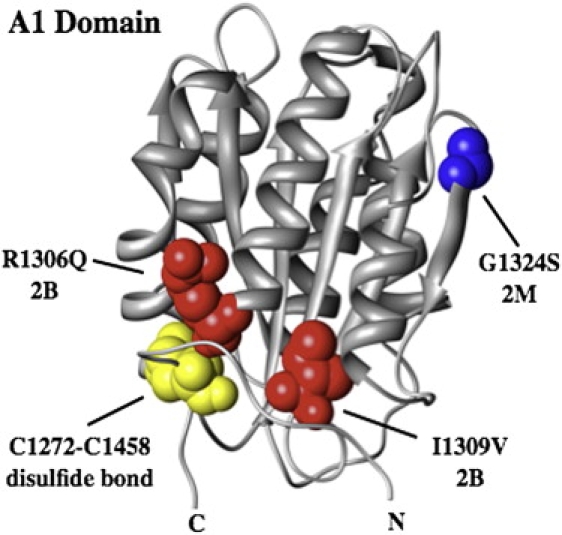
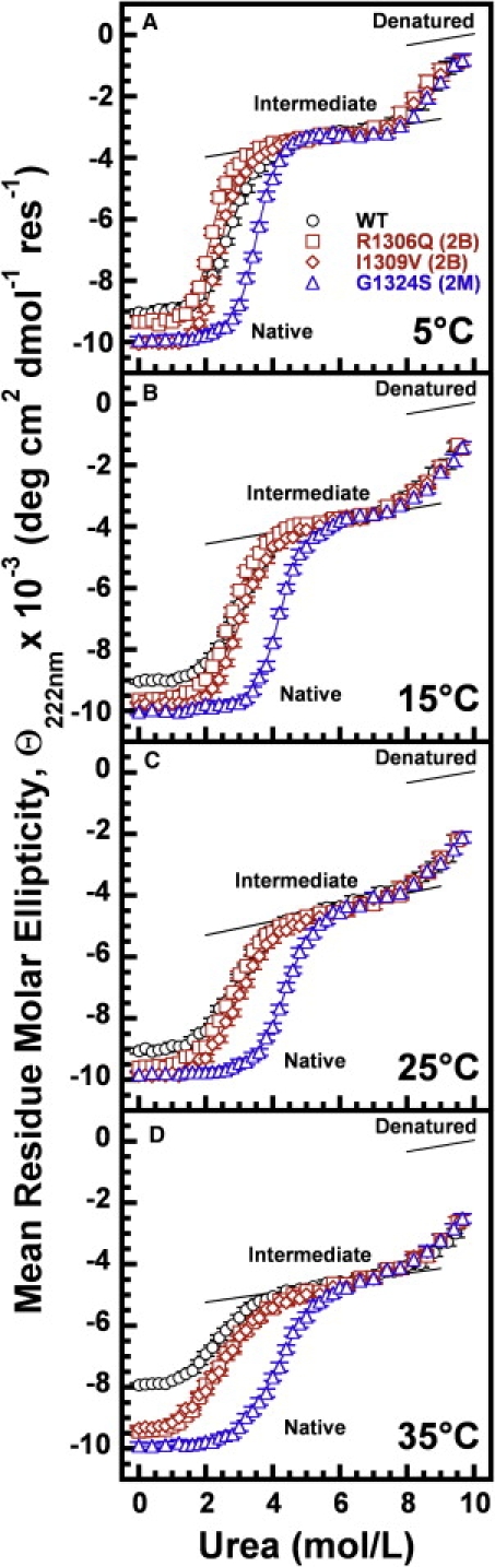
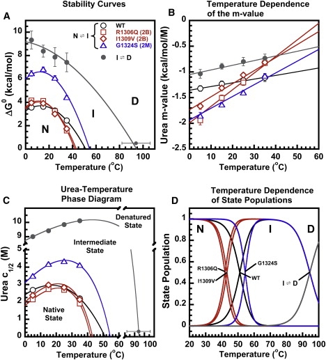
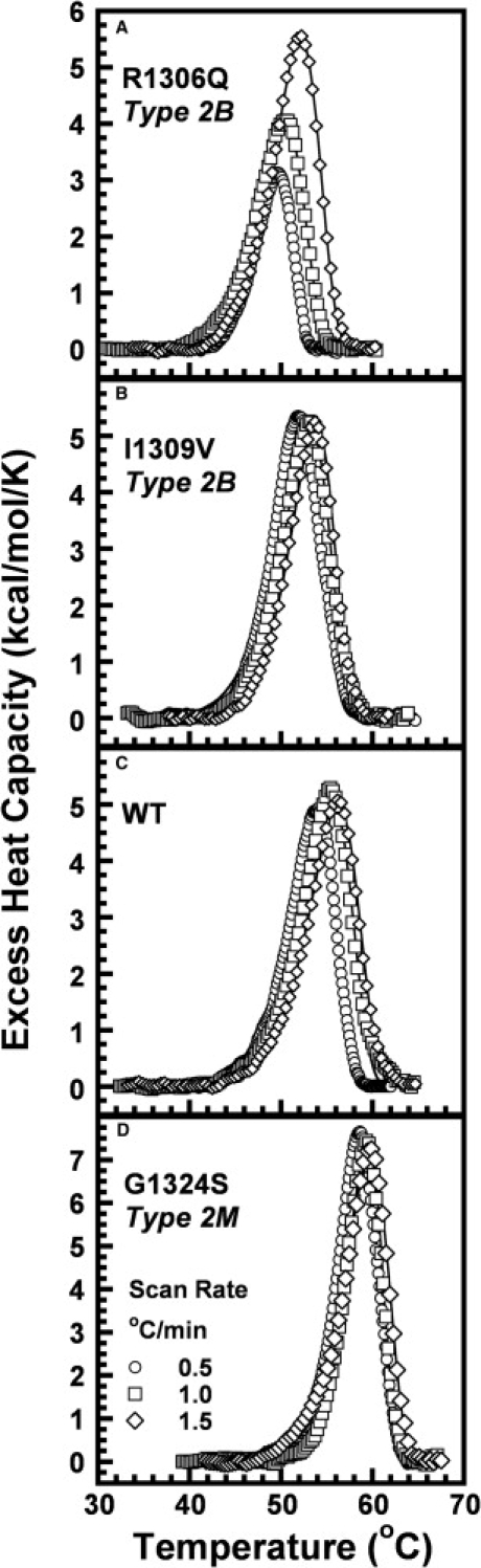
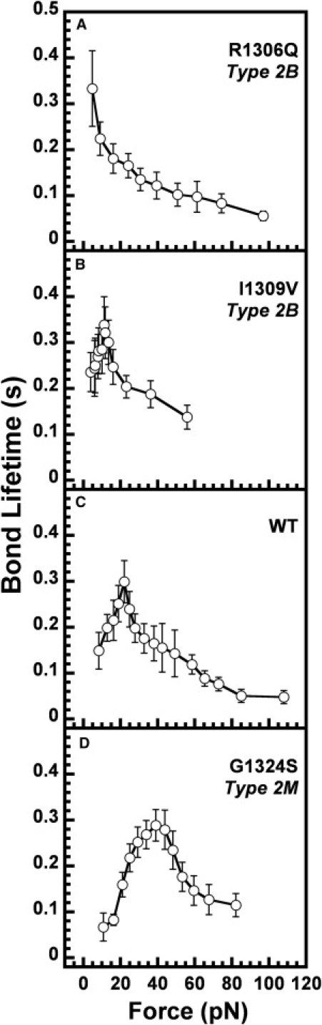
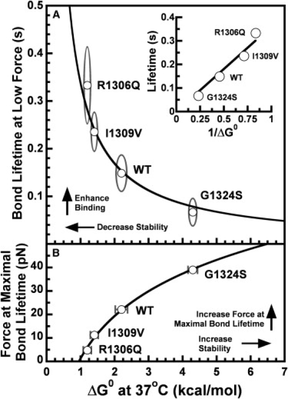
In circulation, plasma glycoprotein von Willebrand Factor plays an important role in hemostasis and in pathological thrombosis under hydrodynamic forces. Mutations in the A1 domain of von Willebrand factor cause the hereditary types 2B and 2M von Willebrand disease that either enhance (2B) or inhibit (2M) the interaction of von Willebrand factor with the platelet receptor glycoprotein Ibalpha. To understand how type 2B and 2M mutations cause clinically opposite phenotypes, we use a combination of protein unfolding thermodynamics and atomic force microscopy to assess the effects of two type 2B mutations (R1306Q and I1309V) and a type 2M mutation (G1324S) on the conformational stability of the A1 domain and the single bond dissociation kinetics of the A1-GPIbalpha interaction. At physiological temperature, the type 2B mutations destabilize the structure of the A1 domain and shift the A1-GPIbalpha catch to slip bonding to lower forces. Conversely, the type 2M mutation stabilizes the structure of the A1 domain and shifts the A1-GPIbalpha catch to slip bonding to higher forces. As a function of increasing A1 domain stability, the bond lifetime at low force decreases and the critical force required for maximal bond lifetime increases. Our results are able to distinguish the clinical phenotypes of these naturally occurring mutations from a thermodynamic and biophysical perspective that provides a quantitative description of the allosteric coupling of A1 conformational stability with the force dependent catch to slip bonding between A1 and GPIbalpha.


Figures

Figure 1
Figure 1
Structure of the A1 domain with the type 2B, R1306Q and I1309V, mutations indicated in red and the type 2M mutation, G1324S, in blue. The disulfide bond is yellow. The structure was drawn using Chimera (http://www.cgl.ucsf.edu/chimera/).
Figure 2
Figure 2
Urea and Guanidine HCl (GndHCl) denaturation of wild-type VWF A1 domain at 25°C monitored by circular dichroism at 222 nm to illustrate the three-state character of the unfolding. Open symbols represent the urea data and solid symbols represent the GndHCl data.
Figure 3
Figure 3
Urea denaturation of wild-type VWF A1 domain (black circles), the type 2B VWD gain-of-function mutations R1306Q (red squares) and I1309V (red diamonds), and the type 2M VWD loss-of-function mutation G1324S (blue triangles) at 5°C, 15°C, 25°C, and 35°C. Lines represent the results of the fit of the data to Eq. S1.
Figure 4
Figure 4
Temperature dependence of the thermodynamic parameters derived from the isothermal urea-induced unfolding of the VWF A1 domain shown in Fig. 3. Symbols are identical to Fig. 3. (A) Thermodynamic stability (ΔG0) of the A1 domain in the absence of urea as a function of temperature defines the stability curve. (B) Temperature dependence of the urea-induced unfolding cooperativity (m-value). (C) Urea-temperature phase diagram. Urea c1/2 versus temperature represents the 50% phase separation line between the native and intermediate states of A1 and between the intermediate and denatured states of A1. (D) Populations of the native, intermediate, and denatured states as a function of temperature. At 37°C, the population of the intermediate for R1306Q is 13%, I1309V is 10%, WT is 3%, and G1324S is <0.1%. The NI transition is represented by open symbols and the ID transition by shaded circles. Data points represent parameters derived from independent fits of the data in Fig. 3 to Eq. S1 and lines represent the results of a global fit of the data in Fig. 2 to Eq. S2.
Figure 5
Figure 5
DSC scans of the VWF A1 domain type 2B VWD gain-of-function mutations R1306Q and I1309V (A and B), wild-type A1 (C), and the type 2M VWD loss-of-function mutation G1324S (D) at scan rates 0.5 K/min (circles), 1.0 K/min (squares), and 1.5 K/min (diamonds). Lines represent the results of the fit of the data to Eq. S5.
Figure 6
Figure 6
A1-GPIbα bond lifetime versus bond force for the type 2B VWD gain-of-function mutations R1306Q and I1309V (A and B), the WT A1 domain (C), and the type 2M VWD loss-of-function mutation G1324S (D). The data in panels A and C were obtained from Yago et al. (14). The data are presented as the mean ± the standard error of the mean.
Figure 7
Figure 7
(A) Bond lifetime determined at low force (near equilibrium) for the interaction between A1 and GPIbα is inversely proportional to the thermodynamic stability of the A1 domain at 37°C. Shaded ellipses represent the error arcs for the experimental error on both the bond lifetime and ΔG0. The fit to Eq. 1 was weighted according to the error on the lifetime measurements. (Inset) Linear plot of bond lifetime versus 1/ΔG0. (B) Dependence of the force required for maximal bond lifetime on the thermodynamic stability of A1. As A1 stability increases, the force for maximal bond lifetime also increases.

Similar articles

Cited by

References

    1. von Willebrand E. Hereditary pseudohemophilia. Finska Läkarssällskapetes Handl. 1926;68:87–112.
    1. Federici A.B., Mannucci P.M. Management of inherited von Willebrand disease in 2007. Ann. Med. 2007;39:346–358. - PubMed
    1. Keeney S., Cumming A.M. The molecular biology of von Willebrand disease. Clin. Lab. Haematol. 2001;23:209–230. - PubMed
    1. Sadler J.E. Biochemistry and genetics of von Willebrand factor. Annu. Rev. Biochem. 1998;67:395–424. - PubMed
    1. Verweij C.L., Quadt R., Briet E., Dubbeldam K., Van Ommen G.B. Genetic linkage of two intragenic restriction fragment length polymorphisms with von Willebrand's disease type IIA. Evidence for a defect in the von Willebrand factor gene. J. Clin. Invest. 1988;81:1116–1121. - PMC - PubMed

Publication types

MeSH terms