. 2009 Jul;17(1):62-74.
doi: 10.1016/j.devcel.2009.05.012.

Sox17 regulates organ lineage segregation of ventral foregut progenitor cells

Affiliations

Sox17 regulates organ lineage segregation of ventral foregut progenitor cells

Jason R Spence et al. Dev Cell. 2009 Jul.

Abstract

The ventral pancreas, biliary system, and liver arise from the posterior ventral foregut, but the cell-intrinsic pathway by which these organ lineages are separated is not known. Here we show that the extrahepatobiliary system shares a common origin with the ventral pancreas and not the liver, as previously thought. These pancreatobiliary progenitor cells coexpress the transcription factors PDX1 and SOX17 at E8.5 and their segregation into a PDX1+ ventral pancreas and a SOX17+ biliary primordium is Sox17-dependent. Deletion of Sox17 at E8.5 results in the loss of biliary structures and ectopic pancreatic tissue in the liver bud and common duct, while Sox17 overexpression suppresses pancreas development and promotes ectopic biliary-like tissue throughout the PDX1+ domain. Restricting SOX17+ biliary progenitor cells to the ventral region of the gut requires the notch effector Hes1. Our results highlight the role of Sox17 and Hes1 in patterning and morphogenetic segregation of ventral foregut lineages.


Figures
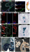
Figure 1
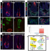
Figure 1. Sox17 and Pdx1 are coexpressed in ventral foregut progenitor cells
A. Sagittal section of an E8.5 embryo showing that Pdx1 (red) and Sox17 (blue) are co-expressed in the ventral foregut in a domain adjacent to the Hhex (green) positive liver primordium. Only a few cells at the boundary between the Pdx1/Sox17 and Hhex domains co-express Pdx1, Sox17 and Hhex (arrows). Right panels show individual channels. The cardiac mesoderm is indicated with dashed lines. The boxed region in the schematic on the right highlights the relative region of the staining and the orientation of the embryo (A – anterior, P – posterior). B. Transverse section of an E9.5 showing that the Sox17/Pdx1 co-expression domain has begun to separate into a ventrally located Sox17-high/Pdx1-low domain a Pdx1-high/Sox17-low domain. Sox17 protein (blue) and Pdx1 protein (red) are shown in separate color channels. C. At E9.5 both the dorsal and ventral foregut are lineage labeled using Pdx1-cre and Rosa26-LacZ (R26R) mice. D. At E10.5 the Pdx1 and Sox17 expression domains resolve into a Pdx1-expressing (red) ventral pancreas (vp) and a Sox17-expressing (green) biliary primordium/gall bladder (pgb). Pdx1 is also expressed in the dorsal pancreas (dp) and midgut (mg). E. The E10.5 biliary primordium/gall bladder comes from a Pdx1-expressing progenitor cell. The gall bladder primordium is negative for the Pdx1 protein but is lineage labeled by Pdx1-cre in Rosa26-LacZ mice. F. Pdx1-cre lineage tracing shows that at E16.5 the duodenum (d), ventral pancreas (vp) dorsal pancreas (dp), gall bladder (gb), collecting duct, common duct and cystic duct are all descendants of a Pdx1 positive precursor. Inset (top) shows a section through the gall bladder lineage labeled with LacZ. Inset (bottom) shows the common duct and cystic ducts lineage labeled with LacZ. G. Pdx1 is not required for normal biliary development. At E16.5, Pdx1 null mice (PdxttTa/tTa) lack a pancreas, but have a gall bladder and cystic duct. Scale bars in A, B and D are 20um.
Figure 2
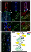
Figure 2. Sox17 is required for development of the biliary primordium and for establishing and/or maintaining organ boundaries between the pancreas and liver
A and B. Pdx1 (red) and Sox17 (blue) expression in the ventral foregut of (A) E9.5 control FoxA3cre;Sox17Fl/+ embryos or (B) FoxA3cre;Sox17Fl/Fl embryos, which show an almost complete loss of Sox17 (blue) expression and a ventral expansion of Pdx1 expression. Dashed lines outline the ventral region of the gut tube where the ventral pancreas (red) and biliary system (blue) are developing. C and D. Pdx1 (green) and Sox17 (red) expression in E10.5 control (C) and FoxA3cre;Sox17Fl/Fl (D) embryos. FoxA3cre;Sox17Fl/Fl embryos entirely lack a biliary/gall bladder primordium. Pdx1 staining (green) marks the dorsal and ventral pancreas in both control and FoxA3cre;Sox17Fl/Fl embryos. E and F. In E10.0 control embryos (E), the ventral Pdx1/Sox17 domains have begun to resolve into a distinct Pdx1 expressing ventral pancreas (red) and a Sox17 expressing biliary primordium (blue). In Pdx1-cre;Sox17Fl/GFP embryos (F) the Sox17-expression domain is replaced by Pdx1-positive epithelium and there is no evidence of a biliary bud. Dashed lines outline the ventral region of the gut tube where the ventral pancreas (red) and biliary system (blue) are developing.G and H. Pdx1 (red), Sox17 (blue) and Prox1 (green) expression in E9.5 control (G) or Foxa3cre;Sox17Fl/Fl (H) embryos. At E9.5 Pdx1 (red) positive cells are almost never seen in the ventral most part of the liver diverticulum (G and G′). However, in FoxA3cre;Sox17Fl/Fl embryos(H and H′) there were many Pdx1 positive cells scattered throughout the liver diverticulum. I. Quantitation of ectopic Pdx1+ cells in the liver diverticulum (Sox17-LOF include FoxA3cre;Sox17Fl/Fl and FoxA3cre;Sox17Fl/GFP embryos). There were 10-fold more Pdx1+ cells in the Sox17-LOF liver bud than in controls (control 0.7 +/− 0.7 cells/liver bud (n=3) vs. Sox17-LOF 7.8 +/− 3 cells/liver bud (n=5), p<0.05). Error bars denote standard error. J. Schematic of Sox17-LOF results. At E9.5, deletion of Sox17 results in loss of biliary cells and an expansion of Pdx1+ cells into the liver bud. By E10.5 Sox17-LOF embryos completely lacked the biliary primordium. The schematic only shows the ventral region of the gut and not the dorsal pancreas. Lines denote the plane of section shown in A-H.
Figure 3
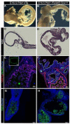
Figure 3. Loss of Sox17 leads to gall bladder agenesis and ectopic pancreas development at e16.5
A and B. As compared to control embryos at e16.5 (A), FoxA3cre;Sox17Fl/Fl embryos (B) completely lack a gall bladder (gb). However, the liver (L), intestine (i), pancreas (p) and stomach (s) appear grossly normal. In (B) the arrow points to a blood vessel (bv) in between the lobes of the liver. A′ and B′ shows a different view of A and B. In this view, the common duct is visible (dashed lines). The boxed area in the inset shows ectopic pancreatic tissue in the common duct. C and D. H&E stained section through the pancreas (P), duodenum (d) and common duct (arrowhead) of a Sox17Fl/Fl control embryo and a FoxA3cre;Sox17Fl/Fl embryo at E16.5. The arrowhead points to the common duct, which has pancreatic tissue budding out of the duct in Sox17-LOF embryos (D). In both controls and Sox17-LOF, the pancreas (P) and duodenum (d) appear normal. C′-C‴ control and D′-D‴FoxA3cre;Sox17Fl/Fl sections through the common duct stained with; the duct specific lectin, DBA (red), insulin plus glucagon (green) and amylase (blue) (C′ and D′); somatostatin (red), insulin (green) and the duct marker, HNF6 (blue) (C″ and D″): or Pdx1 (red), glucagon (green) and HNF6 (blue) (C‴ and D‴). Sox17-LOF ducts have ectopic pancreatic tissue that expresses both exocrine and endocrine markers.
Figure 4
Figure 4. Sox17 suppresses pancreas development and ventralizes the gut
A. Ectopic Sox17 expression in the Pdx1 domain using the Pdx1tTA tetracycline transactivator system. In E10.5 Pdx1tTA;tetO-Sox17 embryos, ectopic Sox17 expression (A′, blue) is seen throughout the Pdx1 expression domain (A, red). The gall bladder primordium expresses endogenous Sox17 (blue), but not Pdx1 (A″, arrowheads). B, D, and F. Control E10.5 embryos expressing Nkx2.2 and Sox17 (B), HNF6 (D), Hhex and Pdx1 (F) protein. C, E and G. Pdx1tTA;tetO-Sox17 embryos at E10.5 expressing Nkx2.2 and Sox17 (C), HNF6 (E), Hhex and Pdx1 (G) protein. In Pdx1tTA;tetO-Sox17 embryos, Nkx2.2 expression in the dorsal and ventral pancreatic buds is dramatically decreased and Hhex expression is expanded dorsally (G;arrowheads). Nkx2.2 is still expressed only in areas where tetO-Sox17 is not activated (C, inset). Inset in F and G shows Pdx1 expression (red). H and I. The transcription factor Ptf1a (green) and Sox17 (blue) in control embryos (H) and in Pdx1tTA;tetO-Sox17 embryos (I). Ptf1a is normally expressed in the presumptive dorsal and ventral pancreas, and but ectopic Ptf1a positive cells are also seen in the middle part of the gut of Sox17-GOF embryos, suggesting a patterning defect. J. Schematic of control vs. Sox17-GOF embryos. In control embryos at E10.5, Nkx2.2 (red) is restricted to the dorsal and ventral pancreas whereas Sox17 (blue) is restricted to the gall bladder primordium and Hhex expression (green) is seen in the liver bud, gall bladder primordium and ventral pancreas. In Sox17-GOF embryos, Sox17 expression is activated throughout the Pdx1 expression domain, which results in failure of the dorsal and ventral pancreas to bud, a decrease in Nkx2.2 expression and a dorsal expansion of Hhex into the gut and presumptive dorsal pancreas.
Figure 5
Figure 5. Sox17 expression is sufficient to induce ectopic ductal tissue, pancreas agenesis and gut malformations at e16.5
A and B. Stomach, duodenum, dorsal and ventral pancreas at E16.5 in control Pdx1tTA (A) and Pdx1tTA;tetO-Sox17 (B) embryos. Sox17-overexpressing embryos develop a large mass of tissue at the border between the stomach and duodenum and have only a pancreatic remnant. C and D. H&E stained sections of control (C) Pdx1tTA and (D) Pdx1tTA;tetO-Sox17 embryos at E16.5 showing the stomach, pylorus, and duodenum. E and F. HNF4α protein (red) is in the duodenum and HNF6 protein (green) is in the developing cystic duct (inset) of Pdx1tTA control (E) embryos at E16.5. In Pdx1tTA;tetO-Sox17 (F) embryos HNF4α and HNF6 are co-expressed in the epithelium of the ectopic mass and HNF6 is expressed in ectopic duct-like structures that are found throughout the mass of tissue. G and H. Villin expression is seen in the normal duodenum of Pdx1tTA control (G) and Pdx1tTA;tetO-Sox17 (H) embryos. However, the ectopic mass of tissue in Pdx1tTA;tetO-Sox17 embryos contains villin positive intestinal epithelium and villin negative epithelium.
Figure 6
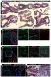
Figure 6. Use of the Tet-regulatible system shows that Sox17 has distinct temporal activities
A through F. Sox17 misexpression between e8 and 10.5 causes organ dysgenesis in the Pdx1 domain. Following Pdx1tTA mediated activation of the tetO-Sox17 transgene at e8.5 we extinguished transgene expression at e9.5 (D), e10.5 (E), and e12.5 (F) by feeding the animals doxycycline (DOX). Control Pdx1tTA embryos developed normally (A, B,C). All Pdx1tTA;tetOSox17 mice (D, E, F) developed a mass and ectopic ducts, indicating that maintaining Sox17 for only a short period of time during development is sufficient to suppress pancreas development and cause mis-patterning of the stomach and duodenum. D′ and D″. Adjacent sections of D were immunostained for the molecular markers HNF6 (green), Pdx1 (blue) and HNF4α (red) (D′) or glucagon (green), insulin (blue) and villin (red) (D″). A large tissue mass developed in these embryos, and HNF6, Pdx1 and HNF4α was seen in ectopic duct-like structures. Ectopic glucagon and insulin expression was seen in outside of the pancreas (P), in between the stomach (S) and duodenum/mass (D). The boxes in D′ and D″ show a magnified view of individual channels in the panels to the right. Arrows and arrowheads point out ectopic glucagon and insulin positive cells, respectively. G and H. Sox17 misexpression in the pancreas from E12.5 results in ectopic ductal tissue at E16.5. E16.5 pancreas from Pdx1tTA control (G) and Pdx1tTA;tetO-Sox17 embryos (H) where tetO-Sox17 was induced at E12.5 (ectopic Sox17 ON at E12.5). E16.5 control embryos show typical cords of Pdx1 (green) positive cells interspersed with DBA (red) positive ducts and surrounded by amylase (blue) positive exocrine pancreas (G). In Pdx1tTA;tetO-Sox17 embryos, cells expressing high levels of Pdx1 are absent and there is a marked increase in DBA positive ducts and a reduction in exocrine pancreas tissue. I and J. Sox17 misexpression in the pancreas from E12.5 results in ectopic ductal tissue in the adult pancreas. Hematoxalin & Eosin staining of adult pancreas from Pdx1tTA controls (I) and Pdx1tTA;tetO-Sox17 embryos (J) where tetO-Sox17 was induced at E12.5 (ectopic Sox17 ON at E12.5). Relative to the control pancreas the Pdx1tTA;tetO-Sox17 pancreas has a dramatic expansion of ductal tissue at the expense of endocrine and exocrine tissue. The inset in J shows a higher magnification of a region that contains numerous ductal structures.
Figure 7
Figure 7. Hes1 and Sox17 may act coordinately to segregate the pancreatobiliary primordia
A and B. Dorsal expansion of Sox17-expressing cells in Hes1−/− embryos at E9.5. Sox17 and Pdx1 protein in Hes1+/− (A) and Hes1−/− (B) embryos at E9.5. In Hes1+/− embryos Sox17 protein (grey) is restricted to the ventral most part of the foregut and Pdx1 protein (red) is highest in the presumptive ventral pancreas. Hes1 protein (green) is found throughout the ventral foregut region and overlaps with both Sox17 and Pdx1. In Hes1−/− embryos (B) Sox17+ cells are expanded dorsally (grey) into the dorsal pancreas (inset, arrows point to ectopic Sox17 cells). See Supplemental Fig. S8 for separated color channels. C and D. The Sox17 and Pdx1 expression domains are disrupted in Hes1−/− embryos at E10.5. Sox17, Hhex and Pdx1 protein in Hes1+/− (C) and Hes1−/− (D) embryos at E10.5. In control embryos (C) Sox17 (blue) and Hhex (green) are expressed in the gall bladder primordium and a Pdx1+ ventral pancreas and gut tube (red). In Hes1−/− embryos (D) the biliary and ventral pancreas are fused (dashed lines) and mostly lack Sox17 (blue) and Pdx1 (red) expression. The Pdx1 expression in D is the gut tube. Scale bars in A-D are 20um. E and F. Hes1 protein is increased in Sox17 gain-of-function embryos. In control e9.5 embryos (E), high Hes1 protein levels are associated with Pdx1/Sox17 co-expressing cells in the ventral foregut. In Pdx1-tTa;tetO-Sox17 embryos, ectopic Sox17 expression in the dorsal bud is associated with elevated Hes1 levels in Pdx1 + cells (E and F, arrowheads). G and H. Hes1 protein is decreased in Sox17 loss-of-function embryos. Compared to control E9.5 embryos, Hes1 protein is only observed in a few cells of the ventral gut of FoxA3cre;Sox17Fl/Fl loss of function embryos (H, arrowheads). For separated Hes1, Pdx1 and Sox17 color channels see supplemental figure 9. Pdx1 protein expression is largely unchanged. Scale bars in A-D are 20um. I. A model derived from Sox17 and Hes1 GOF and LOF data showing how Sox17 and Hes1 might be part of a regulatory loop.

Similar articles

Cited by

References

    1. Ashraf A, Abdullatif H, Hardin W, Moates JM. Unusual case of neonatal diabetes mellitus due to congenital pancreas agenesis. Pediatric Diabetes. 2005;6:239–243. - PubMed
    1. Bhushan A, Itoh N, Kato S, Thiery JP, Czernichow P, Bellusci S, Scharfmann R. Fgf10 is essential for maintaining the proliferative capacity of epithelial progenitor cells during early pancreatic organogenesis. Development. 2001;128:5109–5117. - PubMed
    1. Bort R, Martinez-Barbera JP, Beddington RS, Zaret KS. Hex homeobox gene-dependent tissue positioning is required for organogenesis of the ventral pancreas. Development. 2004;131:797–806. - PubMed
    1. Bort R, Signore M, Tremblay K, Martinez Barbera JP, Zaret KS. Hex homeobox gene controls the transition of the endoderm to a pseudostratified, cell emergent epithelium for liver bud development. Developmental Biology. 2006;290:44–56. - PubMed
    1. Braunstein EM, Qiao XT, Madison B, Pinson K, Dunbar L, Gumucio DL. Villin: A marker for development of the epithelial pyloric border. Dev Dyn. 2002;224:90–102. - PubMed

Publication types

MeSH terms