

Mitral valve hemodynamics after repair of acute posterior leaflet prolapse: quadrangular resection versus triangular resection versus neochordoplasty
- PMID: 19619772
- PMCID: PMC4375960
- DOI: 10.1016/j.jtcvs.2009.01.031
Mitral valve hemodynamics after repair of acute posterior leaflet prolapse: quadrangular resection versus triangular resection versus neochordoplasty
Abstract
Objective: Leaflet prolapse resulting from acute chordal rupture is one presentation of fibroelastic deficiency that is associated with minimal leaflet changes in the prolapsing segment. Minimizing resection and preserving leaflet tissue may be an optimal surgical strategy. We examined the importance of the leaflet preservation concept by comparing resective and nonresective surgical procedures in practice today.
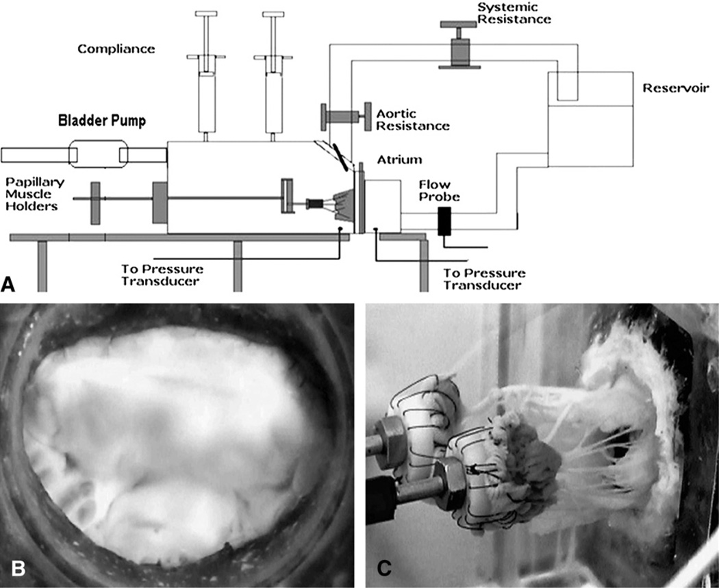
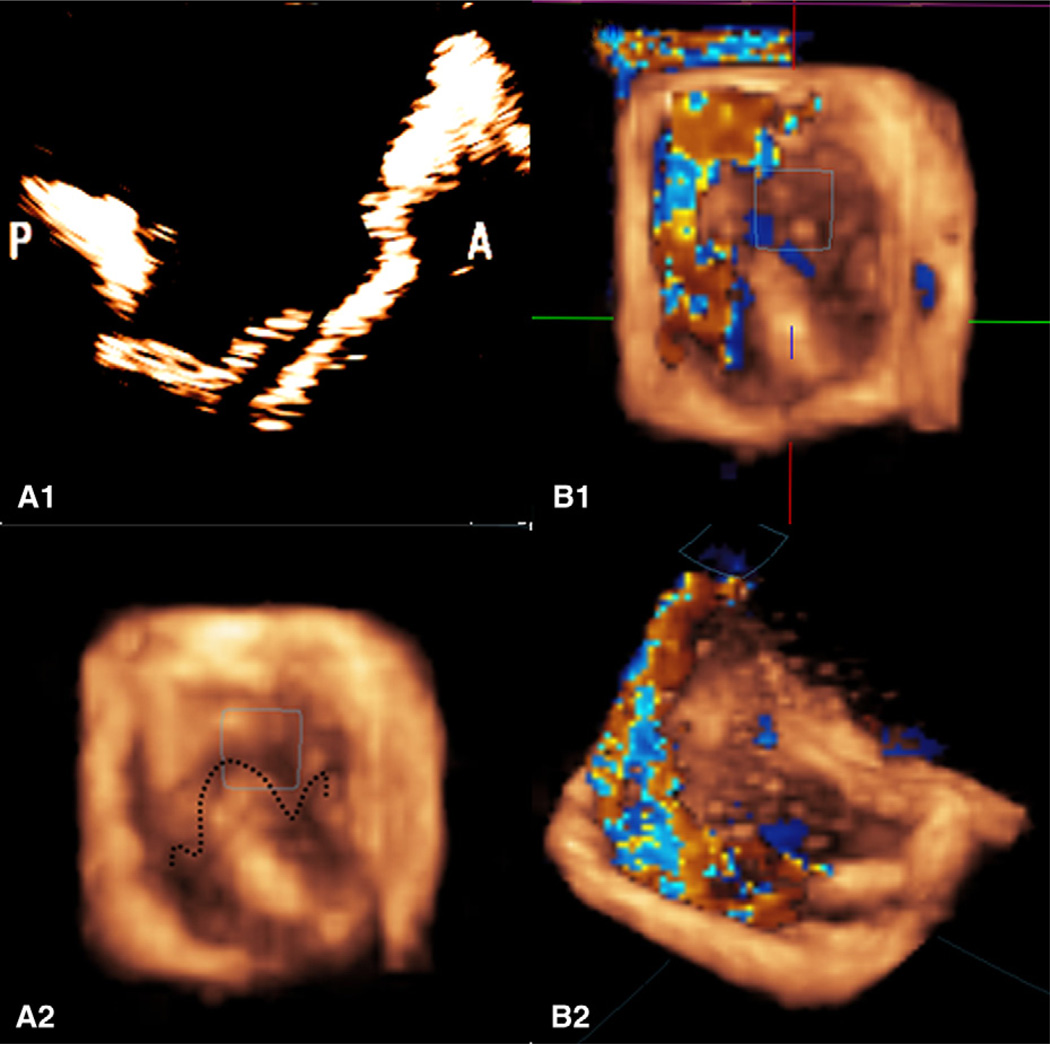
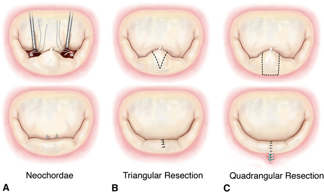
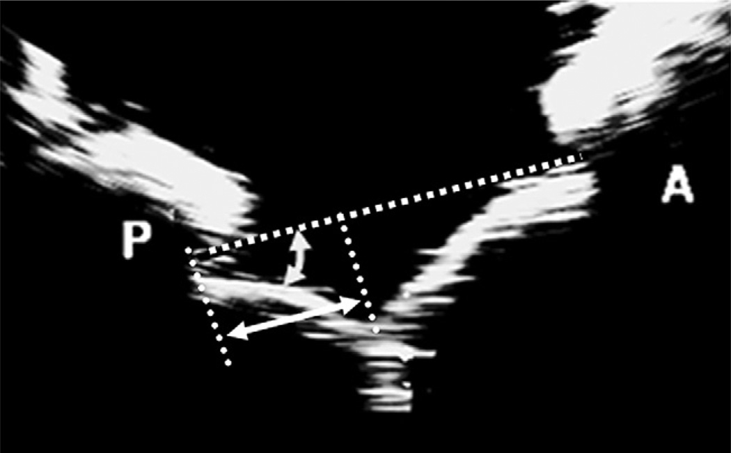
Methods: Eight porcine mitral valves were evaluated in an in vitro heart simulator before surgical manipulation. Mitral regurgitation was created in these valves by transecting the posterior marginal chordae resulting in severe P2 prolapse. After confirmation of mitral regurgiation via regurgitant flow measurement (mL/beat), regurgitation was corrected by three repairs: neochordoplasty with polytetrafluoroethylene sutures (Gore-Tex; W. L. Gore & Associates, Inc, Flagstaff, Ariz), triangular resection, and quadrangular resection with annular compression. Postrepair valve hemodynamics were quantified under pulsatile conditions of 120 mm Hg peak transmitral pressure and 5 L/min cardiac output at 70 beats/min. Furthermore, hemodynamic, geometric, and echocardiographic indices were measured.
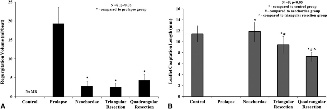
Results: Transecting the marginal chordae resulted in severe P2 prolapse and significant mitral regurgiation (19.3 +/- 4.3 mL/beat). Regurgitant volume was significantly reduced after any of the three surgical approaches (quadrangular, 4.38 +/- 1.6 mL/beat; triangular, 2.56 +/- 1.0 mL/beat; neochordal, 2.86 +/- 1.24 mL/beat). In comparison with the baseline normal valves, leaflet coaptation length and posterior leaflet mobility were significantly reduced in the quadrangular resection group, whereas they were partially restored in the triangular resection and fully preserved in the neochordoplasty group.
Conclusions: Although the three repair procedures are hemodynamically comparable, valve function and leaflet kinematics were significantly better after a nonresection or limited resective correction of leaflet prolapse in this experimental model of acute chordal rupture with otherwise normal leaflet geometry.
Figures
Similar articles
-
Hemodynamic comparison of mitral valve repair: techniques for a flail anterior leaflet.J Heart Valve Dis. 2014 Mar;23(2):171-6. J Heart Valve Dis. 2014. PMID: 25076547
-
Comparison of artificial neochordae and native chordal transfer in the repair of a flail posterior mitral leaflet: an experimental study.Ann Thorac Surg. 2013 Feb;95(2):629-33. doi: 10.1016/j.athoracsur.2012.09.055. Epub 2013 Jan 3. Ann Thorac Surg. 2013. PMID: 23291143 Free PMC article.
-
Chordal replacement versus quadrangular resection for repair of isolated posterior mitral leaflet prolapse.Ann Thorac Surg. 2010 Apr;89(4):1163-70; discussion 1170. doi: 10.1016/j.athoracsur.2009.12.057. Ann Thorac Surg. 2010. PMID: 20338326
-
Is an anterior mitral leaflet prolapse still a challenge?Arch Cardiovasc Dis. 2010 Mar;103(3):192-5. doi: 10.1016/j.acvd.2009.12.002. Epub 2010 Feb 18. Arch Cardiovasc Dis. 2010. PMID: 20417451 Review.
-
Mitral valve repair of isolated posterior leaflet prolapse: resect or respect?Interact Cardiovasc Thorac Surg. 2014 Dec;19(6):1027-35. doi: 10.1093/icvts/ivu279. Epub 2014 Sep 3. Interact Cardiovasc Thorac Surg. 2014. PMID: 25185568 Review.
Cited by
-
Novel Automated Suturing Technology for Minimally Invasive Mitral Chord Implantation: A Preclinical Evaluation Study.Innovations (Phila). 2022 Nov-Dec;17(6):506-512. doi: 10.1177/15569845221133381. Epub 2022 Nov 29. Innovations (Phila). 2022. PMID: 36447382 Free PMC article.
-
Posterior ventricular anchoring neochordal repair of degenerative mitral regurgitation efficiently remodels and repositions posterior leaflet prolapse.Eur J Cardiothorac Surg. 2013 Sep;44(3):485-9; discussion 489. doi: 10.1093/ejcts/ezt092. Epub 2013 Feb 28. Eur J Cardiothorac Surg. 2013. PMID: 23449863 Free PMC article.
-
Left Ventricular Thinning and Distension in Pig Hearts as a Reproducible Ex Vivo Model of Functional Mitral Regurgitation.ASAIO J. 2020 Sep/Oct;66(9):1016-1024. doi: 10.1097/MAT.0000000000001145. ASAIO J. 2020. PMID: 32870609 Free PMC article.
-
Intraoperative transesophageal echocardiography following mitral valve repair: a systematic review.Braz J Anesthesiol. 2022 May-Jun;72(3):379-397. doi: 10.1016/j.bjane.2022.03.002. Epub 2022 Mar 14. Braz J Anesthesiol. 2022. PMID: 35301024 Free PMC article.
-
Commentary: Mitral valve repair using adjustable posterior leaflet neochords.JTCVS Tech. 2020 Feb 20;2:56-57. doi: 10.1016/j.xjtc.2020.01.022. eCollection 2020 Jun. JTCVS Tech. 2020. PMID: 34317751 Free PMC article. No abstract available.
References
-
- Carpentier A. Cardiac valve surgery—the “French correction.”. J Thorac Cardiovasc Surg. 1983;86:323–337. - PubMed
-
- Anyanwu AC, Adams DH. Etiologic classification of degenerative mitral valve disease: Barlow’s disease and fibroelastic deficiency. Semin Thorac Cardiovasc Surg. 2007;19:90–96. - PubMed
-
- Fornes P, Heudes D, Fuzellier JF, Tixier D, Bruneval P, Carpentier A. Correlation between clinical and histologic patterns of degenerative mitral valve insufficiency: a histomorphometric study of 130 excised segments. Cardiovasc Pathol. 1999;8:81–92. - PubMed
-
- He S, Fontaine AA, Schwammenthal E, Yoganathan AP, Levine RA. Integrated mechanism for functional mitral regurgitation: leaflet restriction versus coapting force: in vitro studies. Circulation. 1997;96:1826–1834. - PubMed
-
- Jimenez JH, Liou SW, Padala M, He Z, Sacks M, Gorman RC, et al. A saddle-shaped annulus reduces systolic strain on the central region of the mitral valve anterior leaflet. J Thorac Cardiovasc Surg. 2007;134:1562–1568. - PubMed
Publication types
MeSH terms
Grants and funding
LinkOut - more resources
Full Text Sources
Medical
