. 2009 Sep 18;284(38):26117-26.
doi: 10.1074/jbc.M109.029355. Epub 2009 Jul 20.

Evidence for the direct interaction of spermine with the inwardly rectifying potassium channel

Affiliations

Evidence for the direct interaction of spermine with the inwardly rectifying potassium channel

Masanori Osawa et al. J Biol Chem. .

Abstract

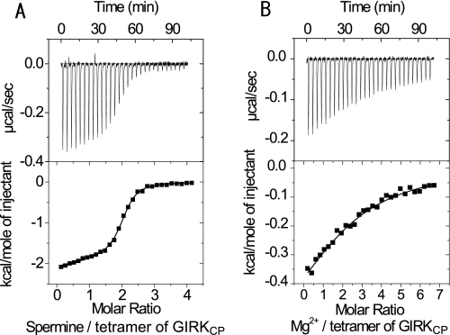
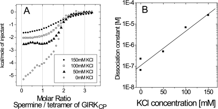
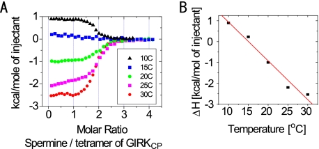
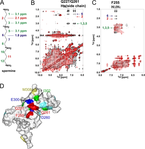
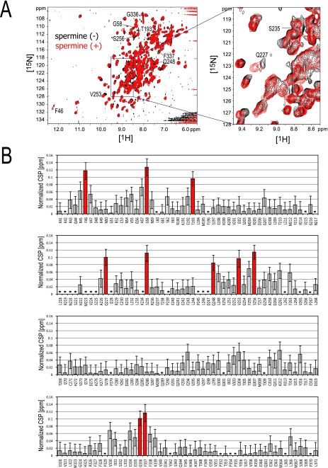
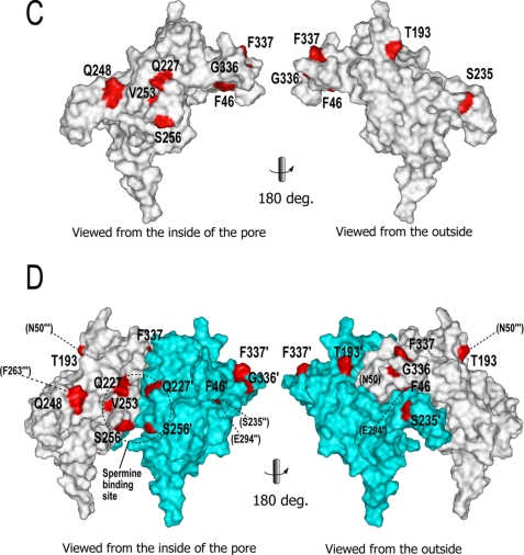
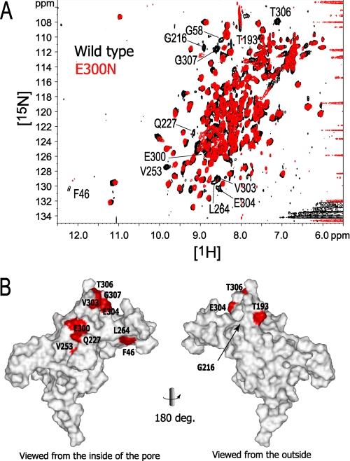
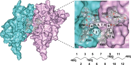
The inwardly rectifying potassium channel (Kir) regulates resting membrane potential, K+ homeostasis, heart rate, and hormone secretion. The outward current is blocked in a voltage-dependent manner, upon the binding of intracellular polyamines or Mg2+ to the transmembrane pore domain. Meanwhile, electrophysiological studies have shown that mutations of several acidic residues in the intracellular regions affected the inward rectification. Although these acidic residues are assumed to bind polyamines, the functional role of the binding of polyamines and Mg2+ to the intracellular regions of Kirs remains unclear. Here, we report thermodynamic and structural studies of the interaction between polyamines and the cytoplasmic pore of mouse Kir3.1/GIRK1, which is gated by binding of G-protein betagamma-subunit (Gbetagamma). ITC analyses showed that two spermine molecules bind to a tetramer of Kir3.1/GIRK1 with a dissociation constant of 26 microM, which is lower than other blockers. NMR analyses revealed that the spermine binding site is Asp-260 and its surrounding area. Small but significant chemical shift perturbations upon spermine binding were observed in the subunit-subunit interface of the tetramer, suggesting that spermine binding alters the relative orientations of the four subunits. Our ITC and NMR results postulated a spermine binding mode, where one spermine molecule bridges two Asp-260 side chains from adjacent subunits, with rearrangement of the subunit orientations. This suggests the functional roles of spermine binding to the cytoplasmic pore: stabilization of the resting state conformation of the channel, and instant translocation to the transmembrane pore upon activation through the Gbetagamma-induced conformational rearrangement.


Figures

FIGURE 1.
FIGURE 1.
Isothermal titration microcalorimetric analyses of the interactions between GIRKCP and spermine or MgCl2. Upper panel, trace of the calorimetric titration of 29 × 10 μl aliquots of spermine (A) and MgCl2 (B) into the cell containing GIRKcp. Lower panel, integrated binding isotherm obtained from the experiment. A, two-site model with the fixed binding stoichiometry of 1 for the binding of the first spermine molecule was applied to fit the data. B, single site model was applied to the MgCl2 binding. Parameters obtained from the best fit (solid line) are summarized in Table 1.
FIGURE 2.
FIGURE 2.
Salt concentration dependence of the GIRKCP-spermine interaction. A, binding isotherms at different concentrations of KCl for the GIRKCP-spermine interaction. B, plot of the dissociation constant value (Kd) against the KCl concentration.
FIGURE 3.
FIGURE 3.
Temperature dependence of the GIRKCP-spermine interaction. A, binding isotherms at different temperatures for the GIRKCP-spermine interaction. B, plot of the enthalpy change (ΔH) against the experimental temperature yielded a ΔCp value of −0.19 kcal/mol·K as the slope of the fitted line.
FIGURE 4.
FIGURE 4.
Intermolecular NOE signals between GIRKCP and spermine. A, chemical structure of spermine, labeled the positions of the methylene groups and their chemical shifts. The chemical shift values are shown for positions 1–3 and 5–6, due to the symmetry of the molecule. B, overlay of the two-dimensional 15N-edited NOESY HSQC spectra for uniformly 2H,15N-labeled GIRKCP in complex with spermine. The spectra for wild type GIRK-spermine and that for the mutant Q227A/Q261A-spermine are shown in black and red, respectively. The assignments of the intermolecular NOE signals are indicated. C, overlay of the NOESY spectra for phenylalanine-selective 1H,15N-labeled GIRKCP in a 2H-background in complex with spermine. The spectra for wild type GIRK-spermine and that for the mutant F255A-spermine are shown in black and red, respectively. The assignments of the intermolecular NOE signals are indicated. D, mapping of the residues with the intermolecular NOEs. The residues with the intermolecular NOEs, Gln-227, Phe-255, and Gln-261 are mapped in red on a surface representation of one of the subunits in the GIRKCP tetramer, which is viewed from the inside of the pore. The amino acid selectively 1H-labeled residues, Ile, Met, and Phe (except for Phe-255), which were used to survey the intermolecular NOE with spermine, are colored green, yellow, and cyan, respectively. The positions of Asp-260 and Glu-300 are also indicated in blue.
FIGURE 5.
FIGURE 5.
Chemical shift perturbation of the backbone NMR signals of GIRKCP upon spermine binding. A, overlay of the TROSY spectra in the presence (red) and absence (black) of spermine. The samples contain 100 μm GIRKCP tetramer and 200 μm spermine (red), or 137 μm GIRKCP (black), respectively. The signals with CSPs larger than 0.08 ppm are labeled. B, CSP upon spermine binding. The x and y axes depict the residues of GIRKCP and the normalized CSP values: {(Δδ1H)2 + (Δδ15N/6.5)2}0.5 (25). The CSPs larger than 0.08 ppm are indicated as red bars. The error bars were calculated based on the digital resolution. Asterisks indicate the residues with no data. C, residues with significant CSPs in B are colored red, with their names on the surface of the single subunit of the crystal structure (PDB code: 1N9P), viewed from the center of the tetramer pore (left) and from the outside (right). Because the coordinates of Gly-58 are missing in the PDB file, Gly-58 was not able to be mapped on the structure. D, mapping of the residues with CSPs (red) on the two adjacent subunits in the GIRKCP tetramer (white and cyan). The residues on the cyan and white subunits are labeled with and without a prime (′). Adjacent residues in other subunits, which are proximal to the residues with CSPs, are shown in parentheses with dotted lines, where double and triple primes indicate the different subunits. The dotted circle depicts the spermine binding site.
FIGURE 5.
FIGURE 5.
Chemical shift perturbation of the backbone NMR signals of GIRKCP upon spermine binding. A, overlay of the TROSY spectra in the presence (red) and absence (black) of spermine. The samples contain 100 μm GIRKCP tetramer and 200 μm spermine (red), or 137 μm GIRKCP (black), respectively. The signals with CSPs larger than 0.08 ppm are labeled. B, CSP upon spermine binding. The x and y axes depict the residues of GIRKCP and the normalized CSP values: {(Δδ1H)2 + (Δδ15N/6.5)2}0.5 (25). The CSPs larger than 0.08 ppm are indicated as red bars. The error bars were calculated based on the digital resolution. Asterisks indicate the residues with no data. C, residues with significant CSPs in B are colored red, with their names on the surface of the single subunit of the crystal structure (PDB code: 1N9P), viewed from the center of the tetramer pore (left) and from the outside (right). Because the coordinates of Gly-58 are missing in the PDB file, Gly-58 was not able to be mapped on the structure. D, mapping of the residues with CSPs (red) on the two adjacent subunits in the GIRKCP tetramer (white and cyan). The residues on the cyan and white subunits are labeled with and without a prime (′). Adjacent residues in other subunits, which are proximal to the residues with CSPs, are shown in parentheses with dotted lines, where double and triple primes indicate the different subunits. The dotted circle depicts the spermine binding site.
FIGURE 6.
FIGURE 6.
The effect of the E300N mutation on the chemical shift. A, The 1H-15N TROSY signals of the E300N mutant that do not overlap with the corresponding signals from the wild-type protein are labeled. B, mapping of the affected residues on the surface of a subunit, viewed from the center of the tetramer pore (left) and from the outside (right). The buried residue (Gly-216) is indicated with an arrow. Because the coordinates of Gly-58 are missing in the PDB file, they were not able to be mapped on the structure.
FIGURE 7.
FIGURE 7.
The effect of the D260N mutation on the chemical shift. A, 1H-15N TROSY signals with chemical shift change larger than the signal line width between the wild type and D260N are labeled. B, mapping of the affected residues on the surface of a subunit, viewed from the center of the tetramer pore (left) and from the outside (right). Buried residues are indicated with an arrow. Because the coordinates of Gly-58 are missing in the PDB file, Gly-58 was not able to be mapped on the structure.
FIGURE 8.
FIGURE 8.
The postulated spermine binding mode. Two adjacent subunits of the tetramer of GIRKCP are shown as the surfaces colored cyan and magenta, in which the spermine binding site is indicated by the dotted rectangle (left). The postulated areas for the spermine atoms in the binding site are illustrated (right). The residues identified in this study are labeled.

References

    1. Bichet D., Haass F. A., Jan L. Y. (2003) Nat. Rev. Neurosci. 4, 957–967 - PubMed
    1. Ficker E., Taglialatela M., Wible B. A., Henley C. M., Brown A. M. (1994) Science 266, 1068–1072 - PubMed
    1. Lopatin A. N., Makhina E. N., Nichols C. G. (1994) Nature 372, 366–369 - PubMed
    1. Lopatin A. N., Makhina E. N., Nichols C. G. (1995) J. Gen. Physiol. 106, 923–955 - PMC - PubMed
    1. Fakler B., Brändle U., Glowatzki E., Weidemann S., Zenner H. P., Ruppersberg J. P. (1995) Cell 80, 149–154 - PubMed

Publication types

MeSH terms

Substances

LinkOut - more resources