. 2009 Sep 11;284(37):24715-24.
doi: 10.1074/jbc.M109.035253. Epub 2009 Jul 20.

Kinetic study of neuropeptide Y (NPY) proteolysis in blood and identification of NPY3-35: a new peptide generated by plasma kallikrein

Affiliations

Kinetic study of neuropeptide Y (NPY) proteolysis in blood and identification of NPY3-35: a new peptide generated by plasma kallikrein

Karim Abid et al. J Biol Chem. .

Abstract

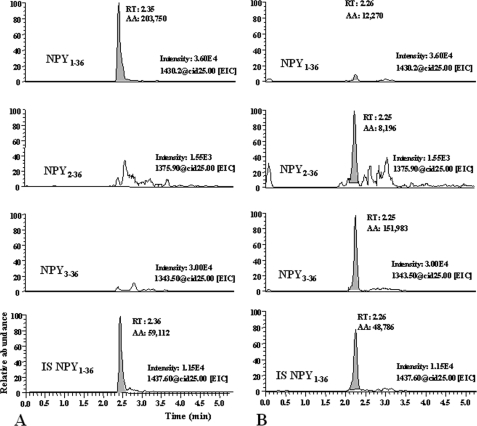
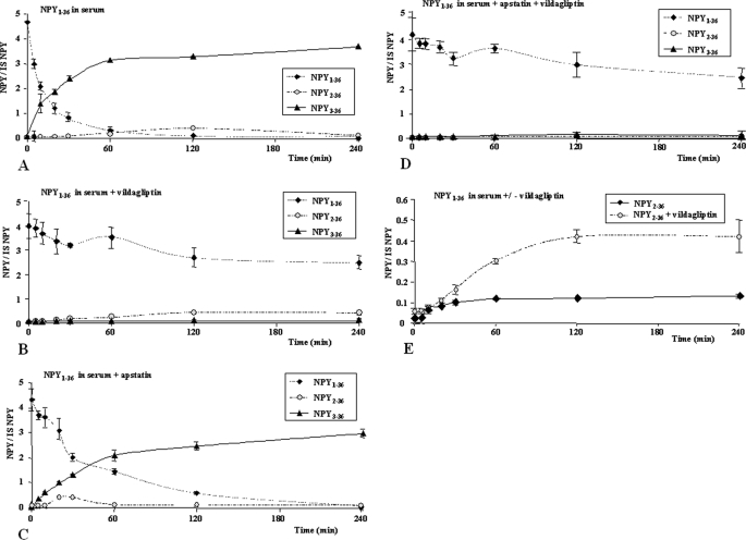
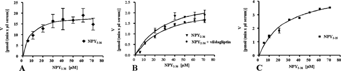
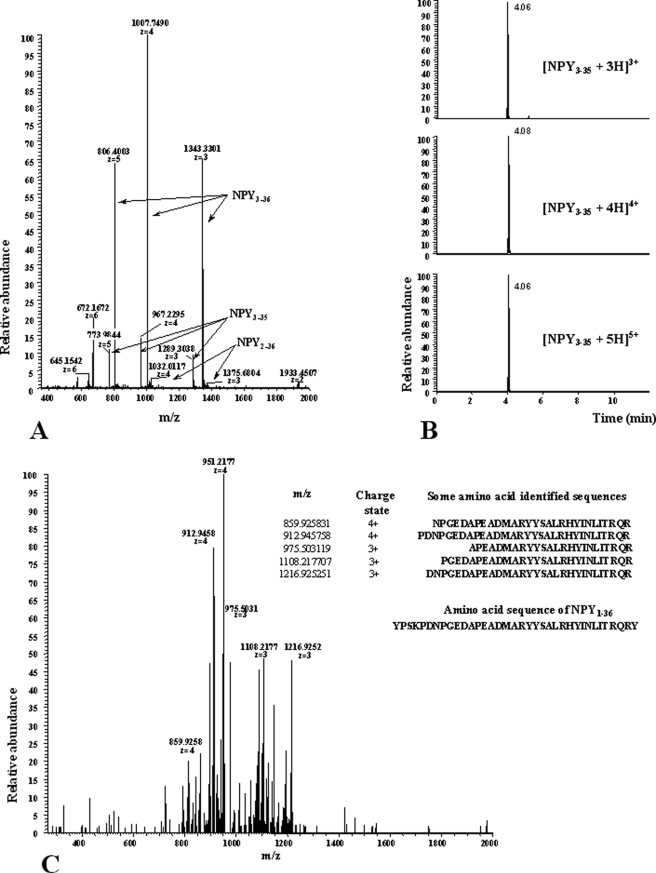
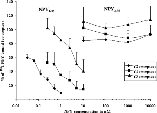
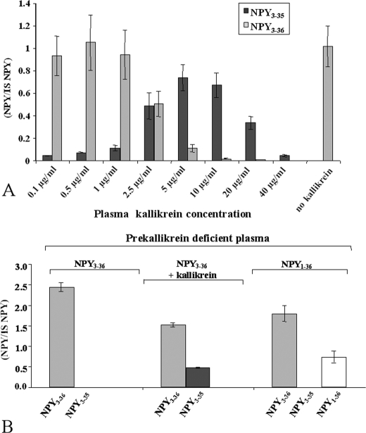
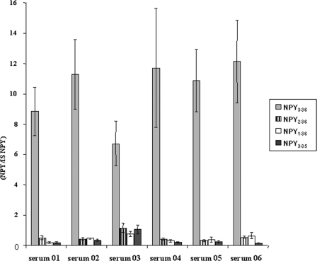
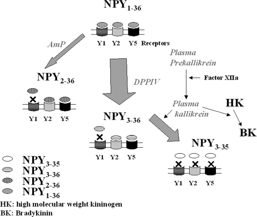
There is little information on how neuropeptide Y (NPY) proteolysis by peptidases occurs in serum, in part because reliable techniques are lacking to distinguish different NPY immunoreactive forms and also because the factors affecting the expression of these enzymes have been poorly studied. In the present study, LC-MS/MS was used to identify and quantify NPY fragments resulting from peptidolytic cleavage of NPY(1-36) upon incubation with human serum. Kinetic studies indicated that NPY(1-36) is rapidly cleaved in serum into 3 main fragments with the following order of efficacy: NPY(3-36) >> NPY(3-35) > NPY(2-36). Trace amounts of additional NPY forms were identified by accurate mass spectrometry. Specific inhibitors of dipeptidyl peptidase IV, kallikrein, and aminopeptidase P prevented the production of NPY(3-36), NPY(3-35), and NPY(2-36), respectively. Plasma kallikrein at physiological concentrations converted NPY(3-36) into NPY(3-35). Receptor binding assays revealed that NPY(3-35) is unable to bind to NPY Y1, Y2, and Y5 receptors; thus NPY(3-35) may represent the major metabolic clearance product of the Y2/Y5 agonist, NPY(3-36).


Figures

FIGURE 1.
FIGURE 1.
Representative LC-MSn recording for NPY1–36, NPY2–36, and NPY3–36 in incubation mix. A, time 0, and B, time 120 min. Fixed scales intensity were chosen at both times to compare left and right chromatograms.
FIGURE 2.
FIGURE 2.
Time course of NPY1–36 degradation in 10% human serum at 37 °C. The values on the y axis represent the ratio digestion products considered versus internal standard (isotopic NPY1–36). A, NPY1–36 degradation in serum without protease inhibitors. B, in the presence of 50 μm vildagliptin. C, in the presence of 50 μm apstatin. D, in the presence of both vildagliptin and apstatin at 50 μm each. E, NPY1–36 digested with or without vildagliptin.
FIGURE 3.
FIGURE 3.
Representation of enzyme kinetics: velocity (v) against concentration (s). Kinetic values are reported in Table 1. A, NPY1–36 was digested at concentrations ranging from 5 to 70 μm (x axis), NPY3–36 production was quantified using LC-MS analysis (porcine NPY1–36 as internal standard). The enzyme velocity is expressed as pmol generated per min and per μl of serum used in the incubation mixture (y axis). B, a similar approach was used for the determination of kinetic constants of AmP toward NPY1–36 without vildagliptin in the incubation mixture. C, determination of the kinetic values of plasma kallikrein toward NPY3–36.
FIGURE 4.
FIGURE 4.
Mass spectrometry characterization of NPY3–35. A, LTQ Orbitrap, full-MS spectra m/z 350–2000; signals of NPY3–35, NPY3–36, and NPY2–36 (charge states +3, +4, and +5 are in bold). B, LC-MS chromatograms for the most abundant isotopes of the triple- (top), fourth- (middle), and fifth-charged ions of NPY3–35 by using a mass window of 2 ppm ([C166H259O54N51S+×H]x+, × = 3, 4, 5). C, MSn spectra of NPY3–35 (charge state +4, m/z 967,2 CID-fragmentation) using a normalized collision energy of 25 and an isolation width of 5 Da; the amino acid sequences listed show the positive hits of the calculated Y-ion type of NPY3–35.
FIGURE 5.
FIGURE 5.
Concentration-response curves of the displacement of 125I-NPY by NPY1–36 and NPY3–35 for the Y1 (■), Y2(♦), and Y5(▴) receptors in SK-N-MC cells (Y1), LN319 cells (Y2), and HEC-1B Y5 cells. Three experiments were performed in duplicate. The percentage of 125I-NPY bound to the receptor, which is caused by the increasing concentrations of NPY1–36, is shown on the y axis. High affinity binding to all NPY receptors was found for NPY1–36. NPY3–35 did not bind to Y1, Y2, or Y5 receptors.
FIGURE 6.
FIGURE 6.
Plasma kallikrein is the carboxypeptidase/esterase enzyme responsible for tyrosinamide removal from NPY. A, purified plasma kallikrein in phosphate-buffered saline is sufficient to cleave NPY3–36 into NPY3–35. NPY3–35 signal was expressed as the ratio versus porcine NPY1–36 used as internal standard; 1 mg of kallikrein corresponds to an activity of 15 units. One unit is defined as the amount of enzyme that hydrolyzes 1 μmol of d-Pro-Phe-Arg-pNA per min at 25 °C, pH 7.8. B, kallikrein-free plasma is unable to degrade NPY3–36 into NPY3–35. Left panel, NPY3–36 added as a substrate in 10% kallikrein-deficient plasma; middle, 0.5 μg/ml purified kallikrein added in the incubation mix; right, NPY1–36 added as a substrate.
FIGURE 7.
FIGURE 7.
Incubation of NPY1–36 in digestion buffer containing serum from 6 different individuals for 1 h. Results are expressed as the ratio of NPY form considered versus porcine standard; experiments were done in triplicate. Serum 01 was the material used to study kinetic parameters of NPY cleavage.
FIGURE 8.
FIGURE 8.
Proposed kinetic model of NPY1–36 cleavage into truncated forms. NPY1–36 is predominantly cleaved into NPY3–36 by DPPIV, and through a slower process by AmP into NPY2–36, resulting for both peptides in a loss of affinity for NPY Y1 receptors. A fraction of NPY3–36 is further degraded by plasma kallikrein into NPY3–35, which do not retain the ability to bind to any NPY receptors. Unknown proteases are likely to be involved in the degradation of NPY1–36, NPY2–36, and NPY3–35 to yield shorter forms, found in trace amounts. See text for further details.

Similar articles

Cited by

References

    1. Wahlestedt C., Edvinsson L., Ekblad E., Håkanson R. (1985) J. Pharmacol. Exp. Ther. 234, 735–741 - PubMed
    1. Walker P., Grouzmann E., Burnier M., Waeber B. (1991) Trends Pharmacol. Sci. 12, 111–115 - PubMed
    1. Abdel-Samad D., Jacques D., Perreault C., Provost C. (2007) Peptides 28, 281–287 - PubMed
    1. Cavadas C., Céfai D., Rosmaninho-Salgado J., Vieira-Coelho M. A., Moura E., Busso N., Pedrazzini T., Grand D., Rotman S., Waeber B., Aubert J. F., Grouzmann E. (2006) Proc. Natl. Acad. Sci. U.S.A. 103, 10497–10502 - PMC - PubMed
    1. Sahu A., Kalra S. P. (1993) Trends Endocrinol. Metab. 4, 217–224 - PubMed

Publication types

MeSH terms