. 2009 Dec;16(4):1313-27.
doi: 10.1677/ERC-09-0082. Epub 2009 Jul 20.

Multiple endocrine neoplasia type 1 knockout mice develop parathyroid, pancreatic, pituitary and adrenal tumours with hypercalcaemia, hypophosphataemia and hypercorticosteronaemia

Affiliations

Multiple endocrine neoplasia type 1 knockout mice develop parathyroid, pancreatic, pituitary and adrenal tumours with hypercalcaemia, hypophosphataemia and hypercorticosteronaemia

Brian Harding et al. Endocr Relat Cancer. 2009 Dec.

Abstract

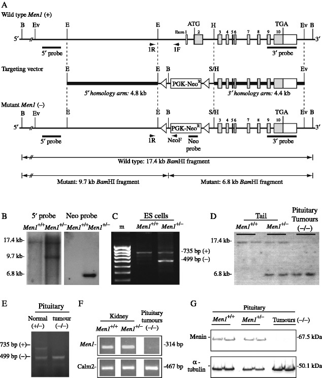
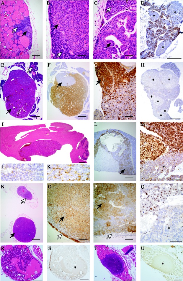
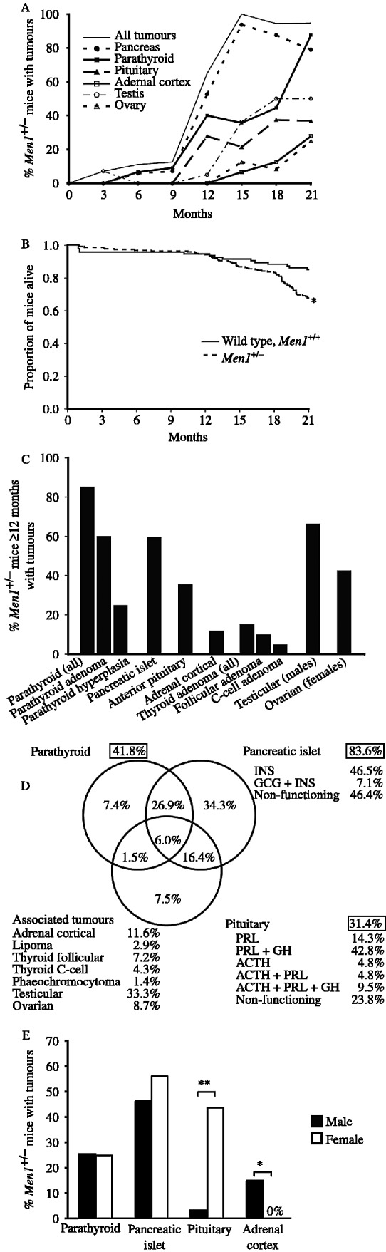
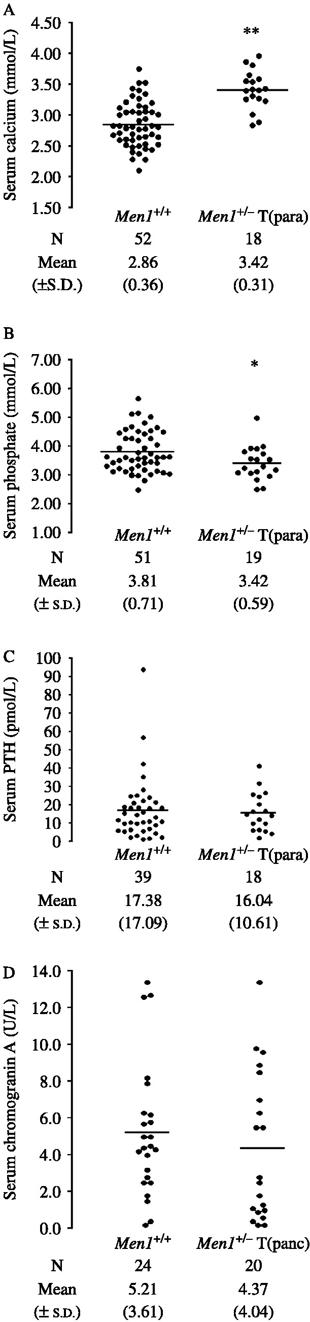
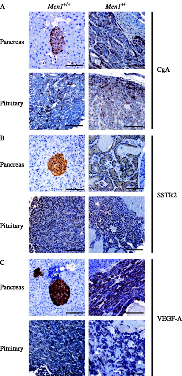
Multiple endocrine neoplasia type 1 (MEN1) is an autosomal dominant disorder characterized in man by parathyroid, pancreatic, pituitary and adrenal tumours. The MEN1 gene encodes a 610-amino acid protein (menin) which is a tumour suppressor. To investigate the in vivo role of menin, we developed a mouse model, by deleting Men1 exons 1 and 2 and investigated this for MEN1-associated tumours and serum abnormalities. Men1(+/-) mice were viable and fertile, and 220 Men1(+/-) and 94 Men1(+/+) mice were studied between the ages of 3 and 21 months. Survival in Men1(+/-) mice was significantly lower than in Men1(+/+) mice (<68% vs >85%, P<0.01). Men1(+/-) mice developed, by 9 months of age, parathyroid hyperplasia, pancreatic tumours which were mostly insulinomas, by 12 months of age, pituitary tumours which were mostly prolactinomas, and by 15 months parathyroid adenomas and adrenal cortical tumours. Loss of heterozygosity and menin expression was demonstrated in the tumours, consistent with a tumour suppressor role for the Men1 gene. Men1(+/-) mice with parathyroid neoplasms were hypercalcaemic and hypophosphataemic, with inappropriately normal serum parathyroid hormone concentrations. Pancreatic and pituitary tumours expressed chromogranin A (CgA), somatostatin receptor type 2 and vascular endothelial growth factor-A. Serum CgA concentrations in Men1(+/-) mice were not elevated. Adrenocortical tumours, which immunostained for 3-beta-hydroxysteroid dehydrogenase, developed in seven Men1(+/-) mice, but resulted in hypercorticosteronaemia in one out of the four mice that were investigated. Thus, these Men1(+/-) mice are representative of MEN1 in man, and will help in investigating molecular mechanisms and treatments for endocrine tumours.


Figures

Figure 1
Figure 1
Targeted disruption of the Men1 gene. (A) Restriction maps of the Men1 gene representing the wild-type (+) and mutant-recombinant (−) alleles. The exons are represented by boxes (filled boxes depict translated regions), and the locations of restriction enzymes sites (BamHI (B), EcoRV (Ev), EcoRI (E), HpaI (H) and SmaI (S)) together with genomic fragments used as 5′, 3′ and neo-cassette (Neo) probes for Southern blot analysis are shown. PCR primers (1R, 1F and NeoF) were designed to facilitate detection of the wild-type (+) and mutant (−) alleles. Exons 1 and 2 together with ∼1.9 kb of the 5′ adjacent region and ∼1.0 kb of intron 2 were disrupted by introduction of the neomycin transferase gene linked to the phosphoglycerate (PGK) promoter (PGK-Neo), in the opposite transcriptional orientation to Men1; the PGK-Neo cassette was flanked by LoxP recognition sequences (open triangles). (B) BamHI Southern blot analysis of genomic DNA extracted from untransfected ES cells (wild-type, Men1+/+) and transfected with Men1 targeting construct (Men1+/−), using the 5′ or Neo probe. Hybridisation with the 5′ probe yielded a 17.4 kb band for the wild-type allele and a 9.7 kb band for the mutant allele, while hybridisation with the Neo probe yielded a 6.8 kb band for the mutant allele, and no signal from wild-type DNA. (C) Genotyping by PCR, utilising primers 1F, 1R and NeoF revealed the presence of the wild-type (735 bp) and mutant (499 bp) alleles from the targeted Men1+/− ES cells, but only the 735 bp from the normal (Men1+/+) ES cells. (D) Southern blot analysis of genomic DNA extracted from mouse tails or pituitary tumours, digested with BamHI and hybridised with the 3′ probe. DNA from Men1+/+ mice showed the expected 17.4 kb band, and the Men1+/− mice showed both the 17.4 and 6.8 kb bands; DNA from the pituitary tumours that developed in Men1+/− mice showed only the mutant 6.8 kb band, consistent with a loss of heterozygosity (LOH) in the tumour. (E) Genotyping by PCR utilising primers 1R, 1F and NeoF revealed the presence of the wild-type (735 bp) and mutant (499 bp) alleles in the normal pituitary of Men1+/− mice, but only the mutant (499 bp) allele from pituitary tumours of these mice, consistent with LOH in the tumours. (F) RT-PCR analysis of normal kidney extracts from Men1+/+ and Men1+/− mice revealed the presence of a Men1 transcript, which was absent in pituitary tumours from Men1+/− mice. Control calmodulin 2 (Calm2) expression is shown. (G) Western blot analysis of normal pituitary extracts from Men1+/+ and Men1+/− mice revealed the expression of menin; however, extracts of pituitary tumours that developed in Men1+/− mice revealed a loss of menin expression. Control immunoblotting for α-tubulin is shown.
Figure 2
Figure 2
Proliferative endocrine lesions in Men1+/− mice. (A–D) Parathyroids, (E–H) pancreas islets, (I–M) pituitary, (N–Q) adrenals and (R–U) thyroids. (A) Nodular parathyroid hyperplasia (black arrow), normal parathyroid (white arrow); (B) parathyroid adenoma (black arrow) and normal parathyroid (white arrow); (C) papillary parathyroid adenoma (black arrow) and adjacent parathyroid (white arrow); (D) parathyroid adenomas showing loss of menin expression (indicated by asterisks); (E) pancreatic islet tumour (black arrow) and a normal sized islet (white arrow); (F) insulin staining of the same tumour showing a non-staining peripheral nodule (black arrow); (G) glucagon-positive nodule of the same tumour (black arrow); (H) multinodular neoplasia of pancreas with loss of menin expression (asterisks); (I) pituitary macroadenoma compressing the overlying cerebrum; (J) anterior pituitary macroadenoma containing prolactin; (K) anterior pituitary macroadenoma containing GH; (L) cystic ACTH containing pars intermedia tumour (arrow); (M) nuclear menin expression was lost in the pituitary adenoma cells, but preserved in adjacent non-neoplastic pituitary cells; (N) unilateral adrenal cortex adenoma (black arrow) and contralateral adrenal cortex hyperplasia (white arrow); (O) this adrenocortical tumour immunostained for 3β-HSD (black arrow) and has compressed residual cortex at the margin (white arrow); (P) phaeochromocytoma showing immunostaining for tyrosine hydroxylase (TH) (black arrow) with compressed cortex unstained with TH at the margin (white arrow); (Q) loss of menin expression in adrenal adenoma (asterisk); (R) thyroid follicular adenoma; (S) serial section showing loss of menin expression in the follicular adenoma (asterisk); (T) thyroid C-cell adenoma; (U) serial section showing loss of menin expression in the C-cell adenoma (asterisk). Scale bars =1000 μm (N), 500 μm (E, F, H, I and R– U), 200 μm (A, L, O and P), 100 μm (D, G and M), 50 μm (B, C and Q).
Figure 3
Figure 3
Tumour occurrence in Men1+/− mice with tumours. (A) Percentage of Men1+/− mice at different ages with tumours. The numbers of Men1+/− mice studied at 3, 6, 9, 12, 15, 18 and 21 months of age were 20, 20, 21, 23, 22, 24 and 19, respectively. The results for the testicular and ovarian tumours are shown as a percentage of the combined total of male and female Men1+/− mice. (B) Kaplan–Meier analysis revealed that survival among the Men1+/− mice (149 out of 220, i.e. <68%) was significantly lower than that in Men1+/+ mice (80 out of 94, i.e. >85%) (*P<0.01, log rank test). Survival in the Men1+/+ and Men1+/− mice was similar up to the age of 12 months, after which survival in the Men1+/− mice began to decrease; this was coincident with the higher frequency of tumour development in the Men1+/− mice that were ≥12 months of age (Fig. 3A and B). (C) Tumour development in Men1+/− mice ≥12 months age, revealed that >85% had parathyroid proliferative abnormalities, which consisted of hyperplasia (∼60%) and adenomas (>25%); >60% had pancreatic islet cell tumours, >35% had anterior pituitary tumours, >10% had adrenal cortical tumours, and >15% had thyroid tumours which consisted of follicular adenomas (>10%) and C-cell adenomas (>5%). Two Men1+/− mice had lipomas, and one had a phaeochromocytoma. Testicular tumours developed in >65% of male Men1+/− mice, and ovarian tumours occurred in >40% of Men1+/− female mice. (D) Schematic representation of 211 tumours that were found in 71 Men1+/− mice. The proportions of Men1+/− mice in which parathyroid, pancreatic or pituitary tumours occurred are shown in the respective boxes. For example, 41.8% of the Men1+/− mice had parathyroid hyperplasia or adenomas. The Venn diagram indicates the proportion of Men1+/− mice with each combination of tumours. For example, 26.9% of Men1+/− mice had both a parathyroid and pancreatic tumour, whereas 34.3% of the Men1+/− mice had a pancreatic tumour only. A similar analysis on the Men1+/− mice aged ≥12 months revealed that >85% had parathyroid abnormalities, >60% had pancreatic islet cell tumours, and >35% had pituitary tumours. In addition, >60% had combined parathyroid and pancreatic, >25% had pancreatic and pituitary tumours; and ∼10% had combined parathyroid, pancreatic islet cell and pituitary tumours; whilst none had combined parathyroid and pituitary tumours. The hormones contained within each of these tumours are indicated: INS, insulin; GCG, glucagon; PRL, prolactin; GH, growth hormone and ACTH, adrenocorticotrophin. (E) Percentage of Men1+/− male and female mice with tumours. P values, *<0.001, **<0.0001.
Figure 4
Figure 4
Serum calcium, phosphate, PTH and chromogranin A concentrations. (A) Serum calcium adjusted for albumin, (B) serum phosphate, and (C) serum PTH concentrations in Men1+/+ and Men1+/− mice with parathyroid hyperplasia or adenomas (Men1+/−T(para)). (D) Serum chromogranin A concentrations in Men1+/+ and Men1+/− mice with histologically proven pancreatic tumours (Men1+/−T(panc)). The results for individual mice are shown. The bars show the mean for each group. P values, *<0.05, **<0.001. The Men1+/−T(para) mice were significantly hypercalcaemic and hypophosphataemic when compared to the Men1+/+ mice. The serum PTH concentrations were similar in both groups, but the occurrence of a serum PTH concentration in the normal range in association with hypercalcaemia is considered to be inappropriate, and consistent with primary hyperparathyroidism (Eastell et al. 2009).
Figure 5
Figure 5
Immunohistochemistry revealing expression of chromogranin A (CgA), somatostatin receptor type 2 (SSTR2), and vascular endothelial growth factor-A (VEGF-A) in pancreatic islets and anterior pituitary tumours of Men1+/− mice. (A) Chromogranin A was present in normal pancreatic islets and anterior pituitary cells of Men1+/+ mice, and in the pancreatic islet cell tumours and anterior pituitary tumours of Men1+/− mice. (B) Somatostatin receptor type 2 was present in Men1+/+ pancreatic islets and anterior pituitary, as well as pancreatic islet and anterior pituitary tumours of Men1+/− mice; (C) VEGF-A expression was observed in Men1+/+ pancreatic islets and anterior pituitary as well as pancreatic islet cell tumours and anterior pituitary tumours of Men1+/− mice.

Similar articles

Cited by

References

    1. Benson L, Ljunghall S, Akerstrom G, Oberg K. Hyperparathyroidism presenting as the first lesion in multiple endocrine neoplasia type 1. American Journal of Medicine. 1987;82:731–737. - PubMed
    1. Bertolino P, Tong WM, Galendo D, Wang ZQ, Zhang CX. Heterozygous Men1 mutant mice develop a range of endocrine tumors mimicking multiple endocrine neoplasia type 1. Molecular Endocrinology. 2003a;17:1880–1892. - PubMed
    1. Bertolino P, Tong WM, Herrera PL, Casse H, Zhang CX, Wang ZQ. Pancreatic beta-cell-specific ablation of the multiple endocrine neoplasia type 1 (MEN1) gene causes full penetrance of insulinoma development in mice. Cancer Research. 2003b;63:4836–4841. - PubMed
    1. Biondi CA, Gartside MG, Waring P, Loffler KA, Stark MS, Magnuson MA, Kay GF, Hayward NK. Conditional inactivation of the MEN1 gene leads to pancreatic and pituitary tumorigenesis but does not affect normal development of these tissues. Molecular and Cellular Biology. 2004;24:3125–3131. - PMC - PubMed
    1. Calender A, Giraud S, Cougard P, Chanson P, Lenoir G, Murat A, Hamon P, Proye C. Multiple endocrine neoplasia type 1 in France: clinical and genetic studies. Journal of Internal Medicine. 1995;238:263–268. - PubMed

Publication types

MeSH terms