. 2009 Oct;29(19):5277-89.
doi: 10.1128/MCB.00001-09. Epub 2009 Jul 20.

Cross talk between receptor guanylyl cyclase C and c-src tyrosine kinase regulates colon cancer cell cytostasis

Affiliations

Cross talk between receptor guanylyl cyclase C and c-src tyrosine kinase regulates colon cancer cell cytostasis

Nirmalya Basu et al. Mol Cell Biol. 2009 Oct.

Abstract

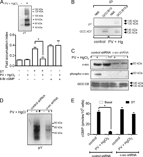
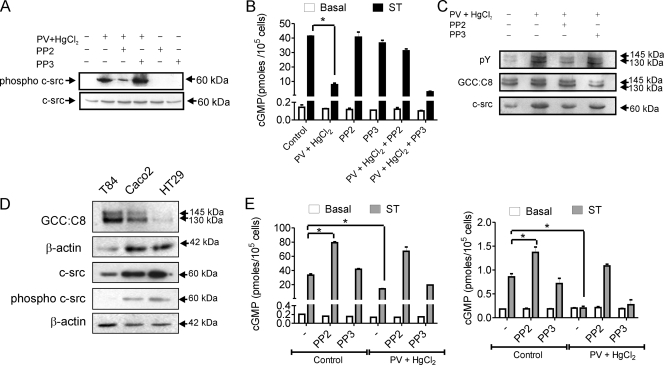
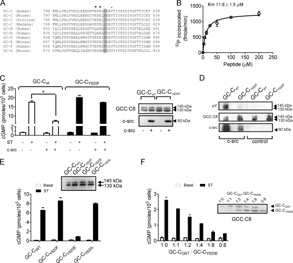
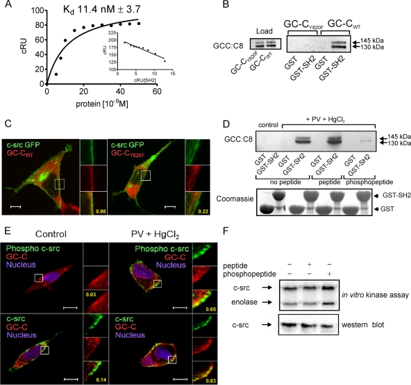
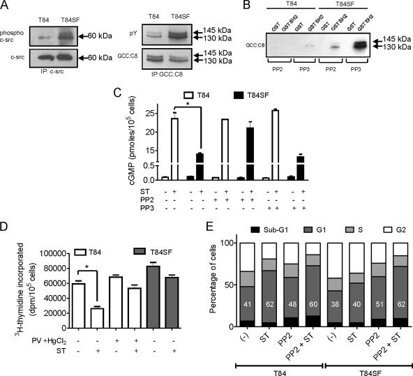
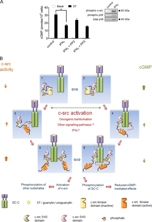
Increased activation of c-src seen in colorectal cancer is an indicator of a poor clinical prognosis, suggesting that identification of downstream effectors of c-src may lead to new avenues of therapy. Guanylyl cyclase C (GC-C) is a receptor for the gastrointestinal hormones guanylin and uroguanylin and the bacterial heat-stable enterotoxin. Though activation of GC-C by its ligands elevates intracellular cyclic GMP (cGMP) levels and inhibits cell proliferation, its persistent expression in colorectal carcinomas and occult metastases makes it a marker for malignancy. We show here that GC-C is a substrate for inhibitory phosphorylation by c-src, resulting in reduced ligand-mediated cGMP production. Consequently, active c-src in colonic cells can overcome GC-C-mediated control of the cell cycle. Furthermore, docking of the c-src SH2 domain to phosphorylated GC-C results in colocalization and further activation of c-src. We therefore propose a novel feed-forward mechanism of activation of c-src that is induced by cross talk between a receptor GC and a tyrosine kinase. Our findings have important implications in understanding the molecular mechanisms involved in the progression and treatment of colorectal cancer.


Figures

FIG. 1.
FIG. 1.
Regulation of GC-C activity by tyrosine phosphorylation in intestinal cells (A) Fluid accumulation was monitored in ligated ileal loop assays following injection of ST (100 nM) or 8-Br-cGMP (5 mM) in the presence or absence of HgCl2 (50 μM) and PV (0.5 mM). The experiment was repeated twice with two loops per treatment, and the values shown represent the mean ± standard error of the mean (*, P < 0.05). Inset, pY Western blot with mucosal scrapings from rat intestinal loops with or without treatment with PV and HgCl2. (B) Mucosal scrapings were prepared from treated and untreated intestinal loops as indicated, and solubilized proteins were allowed to interact with normal mouse immunoglobulin G (NMI) or GCC:B10 monoclonal antibody. Immunoprecipitates (IP) were subjected to Western blot analysis with monoclonal antibody GCC:4D7 and pY antibodies. (C) T84 cells were infected with lentiviruses encoding control or c-src shRNA. Subsequently cells were treated with HgCl2 and PV and harvested, and Western blotting was performed with total c-src, phospho-c-src, and GCC:C8 antibodies. The data shown are representative of experiments repeated thrice. (D) Lysates were prepared from T84 cells infected with lentivirus encoding either control or c-src shRNA following treatment with PV and HgCl2 or medium alone. Lysates were subjected to Western blotting with pY antibodies. The data shown are representative of experiments repeated thrice. (E) T84 cells were infected with lentivirus encoding control or c-src shRNA. Cells were then treated as indicated, following which ST (100 nM) was added and intracellular cGMP produced measured by radioimmunoassay. Values shown represent the mean ± standard error of the mean of duplicate determinations in experiments repeated thrice (*, P < 0.01).
FIG. 2.
FIG. 2.
Activation of c-src in intestinal cells regulates ligand-mediated cGMP production by GC-C, (A) T84 cells were treated as indicated and lysates prepared from cells subjected to Western blot analysis to monitor levels of active c-src (phospho-c-src antibody) and total c-src. The data shown are representative of experiments repeated thrice. (B) T84 cells were treated with HgCl2 and PV following PP2 or PP3 pretreatment for 30 min. cGMP production on addition of ST (100 nM) was measured by radioimmunoassay. Values shown represent the mean ± standard error of the mean of duplicate determinations in experiments repeated thrice (*, P < 0.01). (C) GC-C was immunoprecipitated from T84 cells following treatments as indicated. Immune complexes were subjected to Western blotting with pY and GCC:C8 monoclonal antibodies. To monitor coimmunoprecipitation of c-src, the blot was stripped and then probed with c-src monoclonal antibody. (D) Western blots with lysates from T84, Caco2, and HT29 cells with GCC:C8 monoclonal antibody. The amount of T84 lysate taken was 4 times lower (as was seen with the β-actin loading control) than that of Caco2 and HT29 because of the high expression of GC-C in T84 cells. For Western blotting performed with phospho-c-src and total c-src, whole-cell lysate protein (50 μg) from the three cell lines was used, with β-actin indicating equal loading of the protein. (E) Caco2 (left panel) or HT29 (right panel) cells were treated as indicated and cGMP production on addition of ST (100 nM) measured by radioimmunoassay. Values shown represent the mean ± standard error of the mean of duplicate determinations in experiments repeated thrice (*, P < 0.05).
FIG. 3.
FIG. 3.
Identification of Tyr820 in GC-C as the site for c-src phosphorylation. (A) Amino acid sequence alignment of a region of human GC-C encompassing the Tyr820 residue along with orthologs of GC-C and other receptor GCs. GC-G and GC-E are pseudogenes in human, and therefore the mouse sequences have been used for alignment. *, acidic residues; •, hydrophobic residues downstream of Tyr820. (B) In vitro kinase assay performed with a peptide encompassing the Tyr820 residue and c-src. Values show the mean ± standard error of the mean of duplicate determinations of experiments repeated thrice. (C) HEK293T cells were cotransfected with the indicated plasmids, and following addition of ST (100 nM), the cGMP produced was measured. Values represent the mean ± standard error of the mean of duplicate determinations with each experiment repeated thrice (*, P < 0.01). Right panel, Western blotting performed with GCC:C8 and c-src monoclonal antibodies using total cell lysates. (D) GC-C was immunoprecipitated with GCC:B10 antibody from cells transfected with the indicated plasmids. Immunoprecipitates were subjected to Western blotting with pY, GCC:C8, and c-src monoclonal antibodies. (E) HEK293T cells were transfected with the indicated plasmids. Cells were treated with ST, and the cGMP produced was measured. Values represent the mean ± standard error of the mean of duplicate determinations of experiments repeated thrice (*, P < 0.01). (F) A plasmid encoding GC-CGST was transfected along with various amounts of the GC-CY820E plasmid, and cells were stimulated with ST at 48 h posttransfection. The inset shows a Western blot using GCC:C8 monoclonal antibody to indicate levels of GC-CGST and GC-CY820E in the transfected cells. Values show the mean ± standard error of the mean of duplicate measurements made with experiments repeated thrice.
FIG. 4.
FIG. 4.
Interaction of GC-C with c-src via the SH2 domain contributes to colocalization of c-src and GC-C in the cell. (A) Surface plasmon resonance analysis of c-src SH2 binding to GC-C Y820 phosphopeptide. Inset, linear transformation of data. The data shown are from single sensogram with the experiment repeated twice. (B) GST-SH2 pulldown experiments were performed with lysates prepared from HEK293T cells transfected with either GC-CWT or GC-CY820F plasmid, along with a c-src plasmid. Proteins bound to the glutathione beads were analyzed by Western blotting with GCC:C8 monoclonal antibody. (C) Localization of GC-CWT or GC-CY820F with c-src GFP in HEK293T cells was observed using GCC:4D7 monoclonal antibody. Scale bar, 10 μm. Numbers indicate Pearson's coefficient for the region indicated in the box. The data shown are representative of experiments repeated thrice. (D) T84 cell lysates were allowed to interact with GST-SH2 in the presence of a peptide encompassing the Tyr820 residue or a peptide including a pY residue. Upper panel, Western blotting performed with GCC:C8 monoclonal antibody of proteins bound to beads. Lower panel, Coomassie blue-stained gel of GST and GST-SH2 proteins taken for pulldown experiments. (E) Immunofluorescence imaging was performed with T84 cells with phospho-c-src antibody and GCC:4D7 monoclonal antibody, or with c-src monoclonal antibody and a rabbit polyclonal antibody to GC-C, followed by incubation with anti-mouse Cy5 antibody and anti-rabbit Alexa 488 antibodies. Scale bar, 10 μm. Numbers indicate Pearson's coefficient for the region indicated in the box. The data shown are representative of experiments repeated thrice. (F) In vitro c-src kinase assay performed with c-src immunoprecipitated from HEK293T cells in the presence or absence of peptide or phosphopeptide. The lower panel shows a Western blot to detect the amount of c-src present in the immune complex used for the kinase assay.
FIG. 5.
FIG. 5.
Active c-src prevents GC-C-mediated cell cycle arrest in T84 cells. (A) c-src and GC-C were immunoprecipitated from T84 and T84SF cells with c-src and GCC:B10 monoclonal antibodies, respectively. Western blotting was performed on immune complexes using phospho-c-src and total c-src antibodies (left panel) and pY and GCC:C8 monoclonal antibodies (right panel). The data shown are representative of experiments repeated twice. (B) GST-SH2 pulldown experiments were performed with lysates from T84 or T84SF cells treated with either PP2 or PP3. GC-C bound to the beads was detected by Western blotting using GCC:C8 monoclonal antibody. (C) T84 and T84SF cells were treated with ST (100 nM) with or without PP2 or PP3 pretreatment, and cGMP produced was measured by radioimmunoassay. Values represent the mean ± standard error of the mean of duplicate determinations with experiments repeated thrice (*, P < 0.05). (D) [methyl-3H]thymidine incorporation was measured in T84 cells pretreated with or without PV and HgCl2 and T84SF cells after 24 h of ST (100 nM) treatment. Values represent the mean ± standard error of the mean of duplicate determinations with experiments repeated thrice (*, P < 0.05). (E) T84 and T84SF cells arrested in the G1 phase were treated with medium containing serum in the presence or absence of ST, with or without PP2; 24 h later, the cells were stained with propidium iodide and analyzed by flow cytometry. Numbers in bars indicate the percentage of cells in the G1 phase, and the data shown are representative data from four independent experiments (*, P < 0.05).
FIG. 6.
FIG. 6.
GC-C and c-src cross talk in the intestinal cell. (A) T84 cells were treated with IFN-γ (100 ng/ml) for 24 h. Cells were then either lysed for Western blot analysis with the indicated antibodies or treated with ST for 15 min, following which cGMP was measured by radioimmunoassay. Values shown represent the mean ± standard error of the mean for duplicate determinations for assays repeated thrice (*, P < 0.05). (B) Model for GC-C and c-src cross talk. In a normal intestinal cell, c-src is inactive (1) and/or associated with GC-C via its SH3 domain (2). Under these conditions, most GC-C molecules are unphosphorylated, and on binding ligand (either ST/guanylin/uroguanylin) they produce large amounts of cGMP. Activating mutants of c-src, dysregulated cellular signaling pathways, or ligands (e.g., IFN-γ) contribute to enhanced activation of c-src in the colonic cell. GC-C is now tyrosine phosphorylated at Tyr820 by either free c-src (3) or associated c-src (4). This phosphorylation reduces cGMP production by GC-C. Phosphorylated GC-C can now recruit more c-src via the SH2 domain and, in turn, further phosphorylate GC-C (5 and 6). Binding of c-src to GC-C might also prevent subsequent dephosphorylation of GC-C by cellular phosphatases (5). With an increase in the number of GC-C molecules that are tyrosine phosphorylated, ligand-mediated cGMP production reduces drastically, effectively abrogating the antiproliferative actions of the ligands of GC-C. The c-src activated by interaction with GC-C can now phosphorylate a number of cellular substrates and modulate cell division, migration, and invasiveness.

Similar articles

Cited by

References

    1. Alessandro, R., A. M. Flugy, D. Russo, G. Stassi, A. De Leo, C. Corrado, G. Alaimo, and G. De Leo. 2005. Identification and phenotypic characterization of a subpopulation of T84 human colon cancer cells, after selection on activated endothelial cells. J. Cell Physiol. 203:261-272. - PubMed
    1. Anders, D. L., T. Blevins, G. Sutton, L. J. Chandler, and J. J. Woodward. 1999. Effects of c-Src tyrosine kinase on ethanol sensitivity of recombinant NMDA receptors expressed in HEK 293 cells. Alcohol Clin. Exp. Res. 23:357-362. - PubMed
    1. Bakre, M. M., Y. Ghanekar, and S. S. Visweswariah. 2000. Homologous desensitization of the human guanylate cyclase C receptor. Cell-specific regulation of catalytic activity. Eur. J. Biochem. 267:179-187. - PubMed
    1. Bakre, M. M., and S. S. Visweswariah. 1997. Dual regulation of heat-stable enterotoxin-mediated cGMP accumulation in T84 cells by receptor desensitization and increased phosphodiesterase activity. FEBS Lett. 408:345-349. - PubMed
    1. Banker, N., B. M. Evers, M. R. Hellmich, and C. M. Townsend, Jr. 1996. The role of Src family kinases in the normal and neoplastic gastrointestinal tract. Surg. Oncol. 5:201-210. - PubMed

MeSH terms

LinkOut - more resources