Comparative Study
. 2009 Aug 1;183(3):1644-56.
doi: 10.4049/jimmunol.0900858.

Incorporation of NKT cell-activating glycolipids enhances immunogenicity and vaccine efficacy of Mycobacterium bovis bacillus Calmette-Guerin

Affiliations
Comparative Study

Incorporation of NKT cell-activating glycolipids enhances immunogenicity and vaccine efficacy of Mycobacterium bovis bacillus Calmette-Guerin

Manjunatha M Venkataswamy et al. J Immunol. .

Abstract

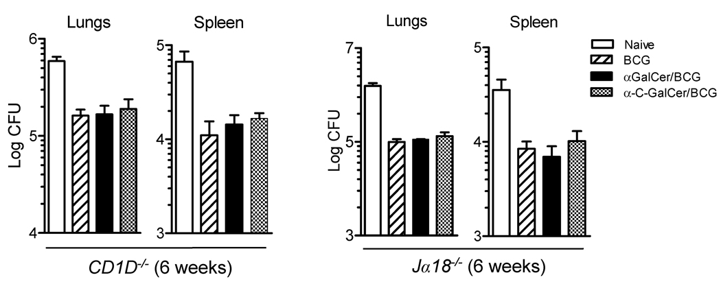
The attenuated strain of Mycobacterium bovis known as bacille Calmette-Guérin (BCG) has been widely used as a vaccine for prevention of disease by Mycobacterium tuberculosis, but with relatively little evidence of success. Recent studies suggest that the failure of BCG may be due to its retention of immune evasion mechanisms that delay or prevent the priming of robust protective cell-mediated immunity. In this study, we describe an approach to enhance the immunogenicity of BCG by incorporating glycolipid activators of CD1d-restricted NKT cells, a conserved T cell subset with the potential to augment many types of immune responses. A method was developed for stably incorporating two forms of the NKT cell activator alpha-galactosylceramide into live BCG organisms, and the impact of this on stimulation of T cell responses and protective antimycobacterial immunity was evaluated. We found that live BCG containing relatively small amounts of incorporated alpha-galactosylceramide retained the ability to robustly activate NKT cells. Compared with immunization with unmodified BCG, the glycolipid-modified BCG stimulated increased maturation of dendritic cells and markedly augmented the priming of Ag-specific CD8(+) T cells responses. These effects were correlated with improved protective effects of vaccination in mice challenged with virulent M. tuberculosis. These results support the view that mycobacteria possess mechanisms to avoid stimulation of CD8(+) T cell responses and that such responses contribute significantly to protective immunity against these pathogens. Our findings raise the possibility of a simple modification of BCG that could yield a more effective vaccine for control of tuberculosis.


Figures

Figure 1
Figure 1. Stable incorporation of aGalCer into live M. bovis BCG
(A) Structures of the iNKT cell activating glycolipids αGalCer and α-C-GalCer. The 14C-labeled form of αGalCer used in this study was identical to that illustrated except that the N-linked acyl chain was 6 carbons shorter (i.e., C20). (B) Solubility of 14C-aGalCer in PBS containing 0.05% Tween 80 or 0.05% tyloxapol, compared to a 2:1 mixture of chloroform and methanol (C:M 2:1) or petroleum ether. The amount of 14C-aGalCer indicated on the X-axis was added to glass vials in C:M 2:1 and the solvent completely evaporated. Y-axis indicates CPM recovered with each solvent when used to redissolve the glycolipid, as determined by β-scintillation counting of an aliquot of the resulting solutions. (C) Growth rate of BCG cultures (monitored by optical density at 600 nm (OD600)) in protein-free Middlebrook 7H9 medium with 0.05% tyloxapol. (D) To determine the optimal concentration of αGalCer for incorporation into live BCG, the bacteria were grown in Middlebrook 7H9 medium with 0.05% tyloxapol containing a range of concentrations of 14C-αGalCer. After growth for 6 days to an OD600 of ~0.8, cultures were harvested and αGalCer incorporation was determined by β-scintillation counting of extensively washed bacteria (% incorporated indicates CPM in the washed bacterial pellet divided by total CPM of 14C-αGalCer added to the culture initially X 100). (E) αGalCer remained intact following incorporation into live BCG. Autoradiograph of a TLC plate is shown. 14C-containing lipids extracted from BCG grown for 8 days (OD600 = 0.7) with 16 µg/ml 14C-αGalCer in Middlebrook 7H9 medium with 0.05% tyloxapol (lane 2) had identical mobility as the 14C-αGalCer reference standard (lane 1). Quantitation of the autoradiographic signals from the TLC plate confirmed that approximately 25% of the radiolabeled glycolipid added to the original culture was stably incorporated into the bacteria as intact αGalCer.
Figure 2
Figure 2. Retention of biologic activity of αGalCer incorporated into BCG
(A) Supernatant levels of IL-2 produced by mouse iNKT cell hybridoma DN3A4-1.2 following culture for 16 hrs with the indicated numbers of autologous BMDC infected at MOI of 10:1 with BCG, or with BCG that was modified by incorporation of either αGalCer (αGalCer/BCG) or α-C-GalCer (α-C-GalCer/BCG). (B) Supernatant levels of IFNγ (left) and IL-4 (right) of naïve C57BL/6 mouse splenocytes (5 × 10⁵) following culture for 48 hrs with autologous BMDC infected at MOI of 10:1 with BCG, αGalCer/BCG or α-C-GalCer/BCG. (C) Hepatic mononuclear cells (4 × 10⁵) from naïve C57BL/6 mice were used as responders, and cultured with autologous BMDC (1 × 10⁵) infected at MOI of 10:1 with the indicated bacteria. Supernatant levels of IFNγ (top) and IL-4 (bottom) were measured after 48 hours. (D) Supernatant levels of IFNγ, TNFα and IL-13 produced by human iNKT cell clone HDE3 following 24 hours of culture with heterologous human monocyte-derived DC infected with BCG, αGalCer/BCG or α-C-GalCer/BCG. (E) In vivo activity of αGalCer incorporated into live BCG. At time 0 hrs, C57BL/6 received a single i.p. injection of either free αGalCer (4.8 nmol in aqueous vehicle), 5 × 10⁶ CFU of unmodified BCG or 5 × 10⁶ CFU of live BCG modified by incorporation of αGalCer (αGalCer/BCG). Serum samples were obtained at 2, 8, 12 and 24 hours post injection, and serum levels of IFNγ, IL-12p70 and IL-4 were determined. For panels A–E, means of triplicate values and standard deviations are shown, and all results shown are representative of at least two independent experiments.
Figure 3
Figure 3. Enhancement of dendritic cell maturation by glycolipid-modified BCG
(A) FACS analysis of indicated markers on CD11c+ cells from spleens of C57BL/6 mice injected i.p. 40 hrs earlier with inert vehicle or with 5 × 10⁶ CFU of live BCG or glycolipid-modified BCG as indicated. The open histograms in the top row indicate staining with isotype matched control antibodies, which gave similar profiles for all four splenocyte preparations. Shaded histograms indicate staining with mAbs specific for MHC class II (I-Ab), CD80, CD86 or CD70 as indicated at the top of each column. The bar graph to the right shows calculated fold increase in median fluorescence intensities for each molecule on CD11c+ cells from spleens of mice immunized with glycolipid-modified BCG compared to mice receiving unmodified BCG. (B) Same analysis as in (A) except for CD11c+ cells in hepatic mononuclear cell suspensions obtained from similarly immunized mice. Results shown are representative of two independent experiments.
Figure 4
Figure 4. Glycolipid incorporation did not alter CD4+ T cell priming or memory responses induced by BCG
(A) Proliferation of transferred p25-specific T cells was observed at similar levels in mice infected with BCG, αGalCer/BCG or α-C-GalCer/BCG BCG. Thy1.1+ B6.PL mice were injected i.v. with CFSE-labeled Thy1.2+ splenocytes from p25TCR-Tg/Rag-1−/− mice, followed by subcutaneous injection either with saline (control), or with 5 × 10⁶ CFU of live bacilli (unmodified BCG, αGalCer/BCG or α-C-GalCer/BCG as indicated). Mice were sacrificed after 7 days, and splenocytes were analyzed by FACS. Dot plots of splenocytes gated for CD4+ and B220 staining are shown for representative mice, and the Thy1.2+ CFSE-labeled populations are indicated in each case within the dashed rectangles. The bar graph on the right indicates the percentage of CD4+ Thy1.2+ cells which retained bright (> FL1 channel 102) CFSE staining. Each bar indicates mean and standard deviation for groups of 3 mice. (B) IFNγ ELISPOT was used to measure recall responses of endogenous CD4+ T cells specific for p25 in spleen cell suspensions from C57BL/6 mice injected intradermally 3 or 8 weeks previously with saline (control) or with 5 × 10⁶ CFU of BCG, αGalCer/ BCG or α-C-GalCer/BCG. All groups receiving unmodified or modified BCG responded similarly. (C) Multiparameter FACS with intracellular staining for cytokines, FoxP3 and CD25 was used to assess the frequencies of multifunctional and regulatory CD4+ T cells (Treg) in cultured splenocytes from animals immunized 8 weeks previously as in (B) and restimulated in vitro with M. tuberculosis (strain H37Rv) sonicate plus anti-CD28 mAb. The graphs show the percentages of total CD4+ T cells simultaneously producing IFNγ, IL-2 and TNFα (multifunctional T cells, left), and the percentages expressing both FoxP3 and CD25 (Treg, right). No significant differences were observed between the groups immunized with BCG versus glycolipid-modified BCG preparations in any of these studies (p > 0.05, ANOVA). All results shown are representative of at least two independent experiments.
Figure 5
Figure 5. Marked enhancement of CD8+ T cell responses by glycolipid-modified BCG
(A) Thy1.1+ B6.PL mice were injected i.v. with CFSE-labeled Thy1.2+ OT-I splenocytes, and infected with the indicated bacteria. CD8+ T cell activation was assessed 7 days after infection by CFSE dilution. Dot plots of splenocytes gated for CD8+ and B220 staining are shown for representative mice, and the Thy1.2+ CFSE-labeled populations are indicated in each case within the dashed rectangles. The bar graph on the right indicates the percentage of CD8+ Thy1.2+ cells which retained bright (> FL1 channel 102) CFSE staining. Each bar indicates the means and standard deviations for groups of 3 mice. (B) ELISPOT assay for IFNγ producing CD8+ T cells specific for the H-2Kb restricted OVA peptide (SIINFEKL) in splenocytes of C57BL/6 mice immunized 3 or 8 weeks previously with saline (control), or with unmodified or glycolipid-modified BCG as indicated. (C) ELISPOT assay for IFNγ producing CD8+ T cells specific for H-2Kd restricted TB10.3/10.4 peptide (GYAGTLQSL) in splenocytes of BALB/c mice immunized 2 weeks previously with saline (control) or with unmodified BCG or αGalCer/BCG as indicated. (D) ELISPOT analysis for IFNγ producing CD8+ T cells specific for H-2Kb presented TB10.3/10.4 epitope (QIMYNPAM) in splenocytes of C57BL/6 mice at 3 weeks post-immunization with unmodified BCG or glycolipid-modified BCG as indicated. Bars indicate responses for animals immunized with BCG with glycolipids physically incorporated (Inc, solid bars), with unmodified BCG plus glycolipids (0.1 µg) injected at a separate site on the opposite flank (Sep; diagonally hatched bars), or with unmodified BCG mixed with glycolipids (0.1 µg) immediately before administration and injected together into the same site (Mix; horizontally hatched bars). (E) IFNγ ELISPOT assay showing CD8+ T cells responses specific for H-2Kb presented TB10.3/10.4 epitope (QIMYNPAM) in spleen cell suspensions from CD1d deficient (CD1D−/−) mice injected intraperitoneally 2 weeks previously with saline (control), or with unmodified or glycolipid-modified BCG as indicated.* p < 0.001; ***, p < 0.0001 (one way ANOVA, Tukey post-hoc test). The vaccine dose was 5 × 10⁶ CFUs for all experiments shown in panels A–E. Results shown are representative of three (A,B) or two separate (C,D,E) independent experiments.
Figure 6
Figure 6. Improved vaccine efficacy against M. tuberculosis challenge with glycolipid-modified BCG
C57BL/6 mice were vaccinated intradermally with saline (naïve, open bars), with 5 × 10⁶ unmodified BCG (diagonally hatched bars) or 5 × 10⁶ glycolipid-modified BCG (αGalCer/BCG, solid bars; α-C-GalCer/BCG, cross hatched bars). Two months later, animals were challenged by aerosol infection with 50–100 CFU of virulent M. tuberculosis (strain H37Rv). Bars show means and SD for CFU of M. tuberculosis in lungs (A) and spleens (B) at 3 and 6 weeks after challenge for groups of 7 mice. π, p < 0.05 (unpaired t test); *, p < 0.05; **, p < 0.007; ***, p < 0.0003 (one way ANOVA, Tukey post-hoc test). Results shown are representative of two independent experiments. (C) Lungs of mice vaccinated and challenged with virulent M. tuberculosis were examined histologically at 6 weeks after challenge. Scale bars: top, 0.5 mm; bottom, 50 µm.
Figure 7
Figure 7. Enhanced vaccine efficacy of glycolipid-modified BCG required iNKT cells
Mice lacking iNKT cells (CD1d−/− or Jα18−/− mice, C57BL/6 background) were vaccinated intradermally with saline (naïve, open bars), with 5 × 10⁶ unmodified BCG (diagonally hatched bars) or 5 × 10⁶ glycolipid-modified BCG (αGalCer/BCG, solid bars; α-C-GalCer/BCG, cross hatched bars). Two months later, animals were challenged by aerosol infection with 50–100 CFU of virulent M. tuberculosis (strain H37Rv). Bars show means and SD for CFU of M. tuberculosis in lungs and spleens 6 weeks after challenge for groups of 4 mice. No significant differences were observed between groups vaccinated with unmodified or glycolipid-modified BCG (p > 0.05, one way ANOVA).

Similar articles

Cited by

References

    1. Dye C, Scheele S, Dolin P, Pathania V, Raviglione MC. Consensus statement. Global burden of tuberculosis: estimated incidence, prevalence, and mortality by country. WHO Global Surveillance and Monitoring Project. JAMA. 1999;282:677–686. - PubMed
    1. Young DB, Perkins MD, Duncan K, Barry CE. Confronting the scientific obstacles to global control of tuberculosis. Journal of Clinical Investigation. 2008;118:1255–1265. - PMC - PubMed
    1. Hoft DF. Tuberculosis vaccine development: goals, immunological design, and evaluation. Lancet. 2008;372:164–175. - PubMed
    1. Andersen P. Tuberculosis vaccines - an update. Nature Reviews Microbiology. 2007;5:484–U16. - PubMed
    1. Hinchey J, Lee S, Jeon BY, Basaraba RJ, Venkataswamy MM, Chen B, Chan J, Braunstein M, Orme IM, Derrick SC, Morris SL, Jacobs WR, Jr, Porcelli SA. Enhanced priming of adaptive immunity by a proapoptotic mutant of Mycobacterium tuberculosis. J.Clin.Invest. 2007;117:2279–2288. - PMC - PubMed

Publication types