. 2009 Oct;77(10):4345-55.
doi: 10.1128/IAI.00542-09. Epub 2009 Jul 20.

Spores as infectious propagules of Cryptococcus neoformans

Affiliations

Spores as infectious propagules of Cryptococcus neoformans

Rajesh Velagapudi et al. Infect Immun. 2009 Oct.

Abstract

Cryptococcus neoformans and Cryptococcus gattii are closely related pathogenic fungi that cause pneumonia and meningitis in both immunocompromised and immunocompetent hosts and are a significant global infectious disease risk. Both species are found in the environment and are acquired via inhalation, leading to an initial pulmonary infection. The infectious propagule is unknown but is hypothesized to be small desiccated yeast cells or spores produced by sexual reproduction (opposite- or same-sex mating). Here we characterize the morphology, germination properties, and virulence of spores. A comparative morphological analysis of hyphae and spores produced by opposite-sex mating, same-sex mating, and self-fertile diploid strains was conducted by scanning electron microscopy, yielding insight into hyphal/basidial morphology and spore size, structure, and surface properties. Spores isolated by microdissection were found to readily germinate even on water agarose medium. Thus, nutritional signals do not appear to be required to stimulate spore germination, and as-yet-unknown environmental factors may normally constrain germination in nature. As few as 500 CFU of a spore-enriched infectious inoculum (approximately 95% spores) of serotype A C. neoformans var. grubii were fully virulent (100% lethal infection) in both a murine inhalation virulence model and the invertebrate model host Galleria mellonella. In contrast to a previous report on C. neoformans var. neoformans, spores of C. neoformans var. grubii were not more infectious than yeast cells. Molecular analysis of isolates recovered from tissues of infected mice (lung, spleen, and brain) provides evidence for infection and dissemination by recombinant spore products. These studies provide a detailed morphological and physiological analysis of the spore and document that spores can serve as infectious propagules.


Figures
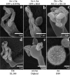
FIG. 1.
FIG. 1.
SEM analysis of C. neoformans spores. Basidia bearing basidiospores from the mating cultures of C. neoformans var. grubii H99α and KN99a (a) and H99α and Bt63a (b), C. neoformans var. neoformans spores from the cross between JEC21α and JEC20a (c), monokaryotic fruiting structures from serotype D α strain XL280 (d), spores from self-filamentous diploid (aAAα) strain KN2B5#19 (e), and control yeast cells from strain H99α (f) were observed by SEM and photographed. Sexual spores from C. neoformans var. grubii are elliptical, with a rough surface that is apparent from the early stages of spore development. Four long intact spore chains on the basidia were commonly seen in the mating cultures from serotypes A and D.
FIG. 2.
FIG. 2.
Comparison of morphologies and spore sizes. Spores produced by mating, fruiting, or diploid self-filamentous C. neoformans strains were analyzed by SEM. A comparative profiling of the spores from H99α and KN99a serotype A mating (a), JEC21α and JEC20a serotype D mating (b), and monokaryotic fruiting of serotype Dα strain XL280 (c) and spores produced by self-filamentous diploid (aAAα) strain KN2B5#19 (d) is shown.
FIG. 3.
FIG. 3.
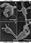
C. neoformans var. gattii sexual spores are elongated. Spores from mating cultures of C. gattii strains are elongated, with a smooth surface, compared to spores from serotypes A and D. Basidia bearing basidiospores that are rod shaped and slightly curved were observed for most of the C. gattii crosses. Different VG types were analyzed by incubating crosses on MS medium, with incubation for 3 to 4 weeks until the spore chains appeared at the edges of the mating colonies. Developing spores at the early stages were round in WM276α matings with B4546a (a), and fully developed, elongated basidiospores were seen in crosses between strains R265α and B4546a (b), NIH312α and B4546a (c), and MMRL2651α and B4546a (d) by SEM.
FIG. 4.
FIG. 4.
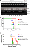
Molecular and mating-type analysis of inocula used for virulence studies. (A) A proportion of the spore inoculum used in the virulence studies was scored for recombination by marker analysis. Mitochondrial (Mito), MATa, and MATα markers were analyzed by PCR using gene-specific primers (see Materials and Methods) and RFLP analysis of one genomic region that perdured in the KN99a/α genome from the original parental strain 8-1, which differs from H99α. (A) Representative sample of the recombination and mating-type analysis conducted. Mating was performed using V8 (pH 7) medium with tester strains JEC20a and JEC21α and was scored for mating type, as shown in Table S1 in the supplemental material. (B and C) Animal studies were performed using 5 (B) or 10 (C) mice, each infected by inhalation of either spores from crosses between strains H99α and KN99a or corresponding yeast cells of H99α and KN99a in an equal (1:1) ratio and/or H99α alone as a control. o/n, overnight.
FIG. 5.
FIG. 5.
Recombinant spore products infect mice and disseminate. CFU from infected mouse tissues (lung, spleen, and brain) were recovered by plating onto Sabouraud dextrose agar medium supplemented with 100 μg/ml chloramphenicol. Molecular and mating-type analyses were performed as described in the legend to Fig. 4. A representative agarose gel of PCR products is shown. Mitochondrial (Mito) DNA inheritance and mating types were determined by PCR using gene-specific primers. Mating types were also determined by mating the isolates with the tester strains (JEC20a and JEC21α) and are shown below the respective panels. (A) Molecular and mating-type analysis of isolates recovered from infected mouse lung tissues, with the corresponding parental strains shown at right. (B and C) Recombinants recovered from infected mouse spleen (B) and recombinants recovered from infected mouse brain (C). A large proportion of recombinants were recovered from infected animal tissues, indicating that spores used as inocula caused infections in mice and disseminated to distant organs. For a complete list of typing, please refer to Tables S1 to S3 in the supplemental material.
FIG. 6.
FIG. 6.
Spores cause lethal infection in Galleria mellonella. Larvae were infected with spores from a cross between strains H99α and KN99a and a 1:1 mixture of H99α and KN99a yeast cells or H99α alone by injection and incubated at 37°C (A) or room temperature (B). Viability was assessed by responsiveness to touch. Survival curves were plotted, and the statistical significance of the survival curves between spores and yeast cells was analyzed (see Results). o/n, overnight.

References

    1. Baker, R. D. 1976. The primary pulmonary lymph node complex of cryptococcosis. Am. J. Clin. Pathol. 65:83-92. - PubMed
    1. Barchiesi, F., M. Cogliati, M. C. Esposto, E. Spreghini, A. M. Schimizzi, B. L. Wickes, G. Scalise, and M. A. Viviani. 2005. Comparative analysis of pathogenicity of Cryptococcus neoformans serotypes A, D and AD in murine cryptococcosis. J. Infect. 51:10-16. - PubMed
    1. Botts, M. R., S. S. Giles, M. A. Gates, T. R. Kozel, and C. M. Hull. 2009. Isolation and characterization of Cryptococcus neoformans spores reveal a critical role for capsule biosynthesis genes in spore biogenesis. Eukaryot. Cell 8:595-605. - PMC - PubMed
    1. Bui, T., X. Lin, R. Malik, J. Heitman, and D. Carter. 2008. Isolates of Cryptococcus neoformans from infected animals reveal genetic exchange in unisexual, α mating type populations. Eukaryot. Cell 7:1771-1780. - PMC - PubMed
    1. Byrnes, E. J., III, R. J. Bildfell, P. L. Dearing, B. A. Valentine, and J. Heitman. 2009. Cryptococcus gattii with bimorphic colony types in a dog in western Oregon: additional evidence for expansion of the Vancouver Island outbreak. J. Vet. Diagn. Investig. 21:133-136. - PMC - PubMed

Publication types