. 2009 Sep 17;114(12):2521-9.
doi: 10.1182/blood-2009-02-205914. Epub 2009 Jul 20.

Activation of endothelial intrinsic NF-{kappa}B pathway impairs protein C anticoagulation mechanism and promotes coagulation in endotoxemic mice

Affiliations

Activation of endothelial intrinsic NF-{kappa}B pathway impairs protein C anticoagulation mechanism and promotes coagulation in endotoxemic mice

Dongmei Song et al. Blood. .

Abstract

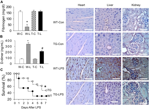
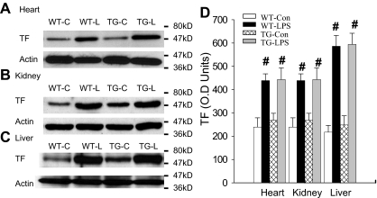
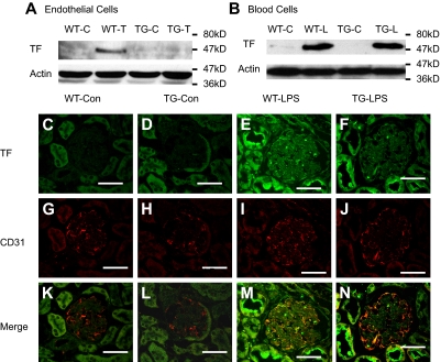
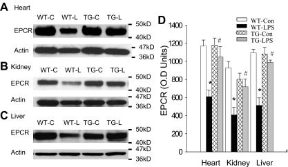
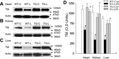
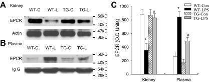
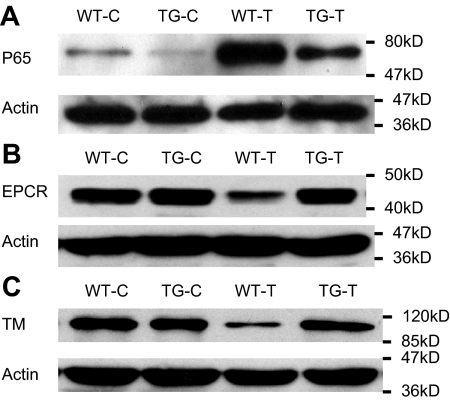
Although the role of systemic activation of the nuclear factor kappaB (NF-kappaB) pathway in septic coagulation has been well documented, little is known about the contribution of endothelial-specific NF-kappaB signaling in this pathologic process. Here, we used transgenic mice that conditionally overexpress a mutant I-kappaBalpha, an inhibitor of NF-kappaB, selectively on endothelium, and their wild-type littermates to define the role of endothelial-specific NF-kappaB in septic coagulation. In wild-type mice, lipopolysaccharide (LPS) challenge (5 mg/kg intraperitoneally) caused markedly increased plasma markers of coagulation, decreased plasma fibrinogen level, and widespread tissue fibrin deposition, which were abrogated by endothelial NF-kappaB blockade in transgenic mice. Endothelial NF-kappaB blockade inhibited tissue factor expression in endothelial cells, but not in leukocytes. Endothelial NF-kappaB blockade did not inhibit LPS-induced tissue factor expression in heart, kidney, and liver. Endothelial NF-kappaB blockade prevented LPS down-regulation of endothelial protein C receptor (EPCR) and thrombomodulin protein expressions, inhibited tissue tumor necrosis factor-alpha converting enzyme activity, reduced EPCR shedding, and restored plasma protein C level. Our data demonstrate that endothelial intrinsic NF-kappaB signaling plays a pivotal role in septic coagulation and suggests a link between endothelial-specific NF-kappaB activation and the impairment of the thrombomodulin-protein C-EPCR anticoagulation pathway.


Figures

Figure 1
Figure 1
Blockade of endothelial NF-κB signaling attenuated coagulation and improved survival. (A-B) Endothelial NF-κB blockade restored plasma fibrinogen level and reduced plasma D-dimer level. Plasma was collected from WT-Con (W-C), WT-LPS (W-L), TG-Con (T-C), and TG-LPS (T-L) groups of mice at 6 hours after saline or LPS injection. Plasma levels of D-dimer and fibrinogen were determined using commercial kits. Data are mean ± SEM of 6 mice. *P < .001 compared with any other group. #P < .001 compared with the WT-LPS group. (C) Endothelial NF-κB blockade improved survival. WT and TG mice were injected with LPS (5 mg/kg intraperitoneally) and followed for 14 days (no further mortality after 7 days). *P < .001, compared with WT mice (log-rank test, 20 mice per group). (D-O) Endothelial NF-κB blockade reduced tissue fibrin deposition. Paraffin-embedded sections were prepared at 6 hours after saline or LPS injection, dewaxed, rehydrated, blocked, reacted with fibrin/fibrinogen-specific antibody, and counterstained with hematoxylin. The dark brown horseradish peroxidase reaction product shows fibrin deposition in microvasculature and arterioles of heart (J), liver (K), and kidney (L) sections from WT-LPS mice. No fibrin deposition was observed in the 3 organs of WT-Con (D-F) and TG-Con (G-I) mice. The dark brown staining was significantly reduced in tissue sections from TG-LPS (M-O) mice. Bar represents 85 μm. Slides were viewed with an Olympus BH2 microscope (Olympus America) using an Splan 40PL lens at 40×/0.70 and Permount mounting medium (Fisher Scientific). Images were acquired using a Nikon DS camera (model DS-U2 cooled), and were processed with Nikon NIS-Elements basic research software and Adobe Photoshop Version 7.0 software.
Figure 2
Figure 2
Blockade of endothelial NF-κB signaling had no effects on tissue levels of TF. (A-C) Western blot photographs showing levels of TF in heart (A), kidney (B), and liver (C) of WT-Con (WT-C), WT-LPS (WT-L), TG-Con (TG-C), and TG-LPS (TG-L) mice at 6 hours after saline or LPS injection. Actin indicates membrane for TF blotting was reblotted with actin antibody. (D) Densitometry quantification of TF bands. Compared with WT-Con and TG-Con, WT-LPS and TG-LPS mice showed significantly increased tissue levels of TF in all 3 organs. Data are mean ± SEM of 5 mice. #P < .01 compared with the WT-Con and TG-Con groups.
Figure 3
Figure 3
Endothelial NF-κB blockade inhibited endothelial TF expression. (A-B) Endothelial NF-κB blockade diminished TNF-α–induced TF protein in ECs (A), but not LPS-induced TF protein in white blood cells (B). WT and TG ECs were treated with Dox (0.5 μg/mL) for 48 hours and then left untreated (WT-C and TG-C) or stimulated with TNF-α (100 ng/mL) for 6 hours (WT-T and TG-T). White blood cells were isolated from WT-Con (WT-C), WT-LPS (WT-L), TG-Con (TG-C), and TG-LPS (TG-L) mice at 6 hours after saline or LPS injection. TF band was not detected in proteins from control cells but induced in TNF-α–stimulated ECs (WT-T) or LPS-stimulated blood cells (WT-L). The TNF-α–induced TF band was prevented in TG ECs (TG-T). The LPS-induced TF band was not affected in TG white blood cells (TG-L). Blots are representative of 3 independent experiments. Actin indicates membrane for TF blotting was reblotted with actin antibody. (C-N) Representative immunofluorescence staining showing endothelial TF expression. Cryosections of kidney were prepared from mice at 6 hours after LPS injection and stained with anti-TF antibody (green) and anti-CD31 (an endothelial-specific marker) antibody (red). Positive staining for TF (E-F) was detected on sections from WT-LPS and TG-LPS mice. TF and CD31 double-positive staining (yellow) localizes TF-expressing ECs (M). TF-expressing ECs (yellow) were not detected in sections from WT-Con and TG-Con mice (K-L), increased in section from WT-LPS mice (M), and significantly reduced on section from TG-LPS mice (N). Bar represents 50 μm. Data are representative of 3 independent experiments. Slides were viewed with a confocal laser-scanning microscope system (FluoView 300-IX; Olympus) using a PLAN APO 60×/1.4 oil objective lens and VECTASHIELD Mounting Medium (Vector Laboratories). Images were processed and analyzed using Image J with colocalization plug-ins that statistically evaluate colocalization using Pearson correlation coefficient (r), which varies from −1 to 1, where 1 equals complete colocalization.
Figure 4
Figure 4
Blockade of endothelial NF-κB signaling reversed LPS-induced EPCR down-regulation. (A-C) Western blot photographs showing levels of EPCR protein in heart (A), kidney (B), and liver (C) of WT-Con (WT-C), WT-LPS (WT-L), TG-Con (TG-C), and TG-LPS (TG-L) mice at 6 hours after saline or LPS injection. Actin indicates membrane for EPCR blotting was reblotted with actin antibody. (D) Densitometry quantification of EPCR bands. Tissue levels of EPCR protein were comparable in all 3 organs between WT-Con and TG-Con mice and were greatly reduced in these organs of WT-LPS mice. Reduction in tissue levels of EPCR protein was not observed in these organs of TG-LPS mice. Data are mean ± SEM of 6 to 8 mice. *P < .001, compared with any other group. #P < .01, compared with the WT-LPS group.
Figure 5
Figure 5
Blockade of endothelial NF-κB signaling prevented LPS-induced TM down-regulation. (A-C) Western blot photographs showing levels of TM protein in heart (A), kidney (B), and liver (C) of WT-Con (WT-C), WT-LPS (WT-L), TG-Con (TG-C), and TG-LPS (TG-L) mice. Actin indicates membrane for TM blotting was reblotted with actin antibody. (D) Densitometry quantification of TM bands. Tissue level of TM protein was high in all 3 organs of WT-Con and TG-Con mice, greatly reduced in organs of WT-LPS mice, and restored in the 3 organs of TG-LPS mice. Data are mean ± SEM of 6 to 8 mice. *P < .001 compared with any other group. #P < .02 compared with the WT-LPS group.
Figure 6
Figure 6
Blockade of endothelial NF-κB signaling reduced endothelial EPCR shedding. (A-B) Western blot photographs comparing tissue (A, kidney) and plasma (B) levels of EPCR protein in same groups of WT-Con (WT-C), WT-LPS (WT-L), TG-Con (TG-C), and TG-LPS (TG-L) mice. Actin and IgG indicate membrane for EPCR blotting was reblotted with actin or IgG antibody. (C) Densitometry quantification of EPCR bands. Compared with WT-Con and TG-Con mice, WT-LPS mice showed a markedly reduced tissue level of EPCR, in parallel with a significantly elevated plasma level of EPCR, indicating EPCR shedding. TG-LPS mice abrogated the LPS-induced reduction in tissue EPCR level and elevation in plasma EPCR level. Data are mean ± SEM of 5 mice. *P < .001 compared with any other group. #P < .001 compared with the WT-LPS group.
Figure 7
Figure 7
NF-κB activation resulted in reduced cellular levels of EPCR and TM in cultured ECs. WT and TG ECs were incubated with Dox (0.5 μg/mL) for 48 hours to induce I-κBαmt expression in TG ECs, and left untreated (WT-C and TG-C) or stimulated with 100 ng/mL TNF-α (WT-T and TG-T) for 1 (for measuring NF-κB activity) or 14 hours (for detecting EPCR and TM proteins). Bolts are representative of 3 experiments. (A) Representative Western blot photograph showing NF-κB activation measured by p65 nuclear translocation. TNF-α markedly activated NF-κB in WT ECs (WT-T), which was significantly inhibited in TG ECs (TG-T). (B) Representative Western blot photograph showing that TNF-α stimulation decreased cellular level of EPCR protein in WT ECs (WT-T), but not in TG ECs (TG-T). (C) Representative Western blot photograph showing that TNF-α stimulation decreased cellular level of TM protein in WT ECs (WT-T), but not in TG ECs (TG-T). Actin indicates membrane for p65, EPCR, or TM blotting was reblotted with actin antibody.

Similar articles

Cited by

References

    1. Martin GS, Mannino DM, Eaton S, Moss M. The epidemiology of sepsis in the United States from 1979 through 2000. N Engl J Med. 2003;348(16):1546–1554. - PubMed
    1. Liu SF, Malik AB. NF-κB activation as a pathologic mechanism of septic shock and inflammation. Am J Physiol Lung Cell Mol Physiol. 2006;290(4):L622–L645. - PubMed
    1. Schouten M, Wiersinga WJ, Levi M, Van Der Poll T. Inflammation, endothelium and coagulation in sepsis. J Leukoc Biol. 2008;83(3):536–545. - PubMed
    1. Oeth PA, Parry GC, Kunsch C, Nantermet P, Rosen CA, Mackman N. Lipopolysaccharide induction of tissue factor gene expression in monocytic cells is mediated by binding of c-Rel/p65 heterodimers to a kappa B-like site. Mol Cell Biol. 2004;14(6):3772–3781. - PMC - PubMed
    1. Mackman N, Brand K, Edgington TS. Lipopolysaccharide-mediated transcriptional activation of the human tissue factor gene in THP-1 monocytic cells requires both activator protein 1 and nuclear factor kappa B binding sites. J Exp Med. 1991;174(6):1517–1526. - PMC - PubMed

Publication types

MeSH terms